Thursday, December 11, 2014

Holiday Foot Care Reminders


With the holidays fast approaching, many individuals may feel the mounting pressure of finding the perfect gift by spending hours at the mall.  The pressure placed on feet dashing from store to store can add up, and in individuals with diabetes can become dangerous.   Even though the holiday of St. Nicholas passed this past week, in which children leave their shoes out to be filled with treats, individuals with diabetes may still want to leave their old shoes out for good and opt for a better, pressure relieving pair.  

High pressure in a healthy foot signals pain, and subsequent alleviation of the inciting pressure by the person moving their foot or changing their shoe to stop the pain from occurring.  In diabetic individuals with peripheral neuropathy, pain and areas of high pressure cannot be felt due to loss of sensation.  This loss of sensation causes of loss of protection to the foot and wounds may develop in areas of abnormally high pressure.  Any deformity that predisposes the foot to rub in a shoe creates an area that is more likely to ulcerate.  Surgery to remove deformities including bunions, hammer toes and bony bumps in the ball of the foot are one of the ways that your podiatrist may remove an area of increased pressure that is likely to develop or has previously developed an ulcer.  Surgery to lengthen the tendon that attaches the calf muscles to the heel bone is also used to lessen pressure on the ball of the foot.  When this tendon is too tight, the heel is lifted and the front of the foot is levered down towards the ground, creating an area of high pressure. 


Along with surgery, regular callus and corn removal by your podiatrist is also very helpful in removing pressure.  Shoes, casting and custom orthotics are all used to better disperse pressure on the foot.  These treatment means are also useful to prevent ulcers from developing or recurring.  By custom molding shoes to the foot, deformities are accommodated for and areas of high pressure are relieved.  Custom orthotics for diabetic patients with neuropathy are made with several layers of material so that any friction that would normally be imparted to the foot is instead absorbed by the orthotic.  Talk to your podiatrist about taking pressure off this holiday season and keeping your feet healthy!


Brian Timm, DPM, FACFAS

Monday, November 24, 2014

National Diabetes Awareness Month


diabetic foot doctor Columbus,neuropathy specialist Columbus OH,diabetic ulcer Columbus OH


For most people, November is the time of year to begin preparing for the holiday season, but November is also National Diabetes Month.  In the United States alone, 25.8 million children and adults have diabetes.  This makes up about 8.3% of the population, and is projected to reach 21% by the year 2050.  There are millions of Americans with undiagnosed diabetes.

Many more who will eat candied apples, canned cranberries, apple and pumpkin pie, and all the turkey and stuffing they can handle; and may likely be precipitating their diabetes and obesity even further without even being aware that they have the signs/symptoms of diabetes in the first place. 

Early warning signs of the disease:
  • Increased thirst
  • Increased hunger (especially after eating)
  • Dry mouth
  • Frequent urination or urine infections
  • Unexplained weight loss (even though you are eating and feel hungry)
  • Fatigue (weak, tired feeling)
  • Blurred vision
  • Headaches

Diabetes can affect the entire body, including the foot and ankle.  Neuropathy is one of the most common complaints of diabetic patients.  Neuropathy results in damage to nerves, specifically the peripheral nerves, or nerves outside of the brain and spinal cord. Peripheral Neuropathy causes a burning or tingling sensation usually on the feet and legs, or on the arms.  This damage to the nerves is caused by excess blood glucose, or high blood sugar.  Over time excess glucose injures the walls of blood vessels that supply the nerves.  As neuropathy worsens, the ability to feel the lower extremity becomes diminished, which makes diabetics more prone to injuries that go unnoticed.  Neuropathy is the leading cause of diabetic foot ulcers.  Diabetic foot ulcers are caused from a combination of neuropathy, trauma, and deformity.  Deformities such as hammertoes, equinus (limited upward bending of the ankle), or bunions in diabetic patients are more problematic than in non-diabetic patients because of the other complications that come with diabetes.  If diabetic foot ulcers are left untreated they may end up getting infected, and even result in amputation.  Diabetic patients often have decreased circulation in the extremities due to hardening of the arteries or vessels, and this may slow healing time, which is why it’s vital for diabetic patients to take extra precautions when it comes to caring for their feet. 


Although diabetes is a life altering disease keeping a tight control on blood glucose levels can minimize complications.  Other ways to protect the feet in individuals with diabetes are by wearing special shoes made for diabetics, examining the feet daily, washing the feet and making sure to dry them completely, avoiding smoking, wearing clean, dry socks that are changed daily, and never walking barefoot.  Management of diabetes is the key to preventing complications, and for those that do not have diabetes the key to prevention is exercise, maintaining a healthy weight, and eating a well balanced diet.

Brian Timm, DPM, FACFAS
Board Certified by the American Board of Foot and Ankle Surgery in Foot and Reconstructive Rearfoot and Ankle surgery

Thursday, November 20, 2014

External fixation here in Naples Florida


External fixation may look very high-tech, and maybe even scary.  However, external fixation has been used in one way or another since almost 2400 years ago.  External fixation techniques were described by Hippocrates, and were used in treating tibia fractures.  External fixation is a minimally invasive technique to reduce displaced fractures and has become a very important part of deformity correction and nonunion and pseudo arthrosis repair as well.

Jean François Malgaigne was one of the many pioneers that made advancements with external fixation devices.  In 1846, Malgaigne used a device that consisted of a clamp and four metal prongs to reduce and stabilize a fracture of the patella, or kneecap.  Following this external fixation device many other similar inventions were used to treat fractures in various locations.  In 1938, Raoul Hoffman made advancements that made external fixation even more useful, and allowed surgeons to place pins into a fracture for stabilization with guidance, while being minimally invasive.  In 1951, Dr. Gavriil A. Ilizarov developed the external fixation device that is still in use today.  Ilizarov’s fixation device consists of a metal frame that encircles the limb, and is attached to underlying bone by pins.  Threaded rods and hinges allow movement of the bone to the correct alignment.  Ilizarov’s external fixator is great because adjustments can be made without opening the fracture site, and the device provides stability. Also it can be converted to internal fixation once the soft tissues have become less swollen. 


Modern day external fixation not only provides stability to a fracture, but can also be used for soft tissue deformities, as well as other bony deformities.  External fixation is preferred when slow correction is required, and even more useful in high risk patients with vascular disease and even open fractures with high risk of infection.  The chance of getting a blood clot is lessened because with external fixation patients can be partial weight bearing, or weight bearing as tolerated following the procedure due to the stability that the fixation provides.  External fixators have been used for other bony deformities, such as Charcot, ankle arthritis, and clubfoot.  External fixation can also be used to lengthen amputated foot and toe stumps.  External fixation has definitely helped many people, but there are still some cons.  Pain and infections are two issues associated with external fixation, and rates vary depending on the extent of the procedure, and the location of the device.  External fixation devices are used by specially trained physicians and all three of the doctors at FFLC are capable of utilizing these various techniques for patients that require such interventions.  


Brian Timm, DPM, FACFAS
Board Certified by the American Board of Foot and Ankle Surgery in Foot and Reconstructive Rearfoot and Ankle surgery

Monday, November 10, 2014

Swimming and your feet


Everyone is starting to return to Florida for the seasonal upgrade in weather.  Fire up the pool warmer, and get ready to swim.

 Swimming is known for being an activity that is less damaging to the joints of the knee, foot and ankle, and can even offer a means of exercise for athletes of other sports with certain foot injuries!
Just about the worst thing a runner can hear is that they are injured and going to have to take time off from running.  Luckily, swimming can help soften this blow by offering an alternative way to get some cardiovascular activity.  Stress fractures are small fracture that occurs from excessive force on normal bone and can force runners to talk a break from running for several weeks.  For the runner with a stress fracture, swimming is often a great alternative that will not bear weight on the injury.  Swimming can also help an athlete ease back into their favorite sport after other serious injuries.  For example, after a peroneal tendon tear, swimming can help to make the transition from being immobilized in a cast or a CAM walker boot to normal running or other weight bearing sports go more gently by allowing the muscles to slowly adapt.  
Swimmers should also be careful to take care to avoid injuries that can occur from intense pool training.  Ankle pain is a common complaint from the repetitive motion the ankles are forced through while swimming.  The Achilles tendon, which is responsible for flexing the foot, may also become irritated from the repetition and develop Achilles tendonitis.  While the Achilles is doing much of the work, the extensor tendons on the top of the foot can also become irritated by being tightly pulled.  By taking care to stretch the feet and ankles properly and always listen to your body when working out, these injuries can be minimized.  Swimmers also need to take extra care to avoid developing warts, Athlete’s foot, and fungal nails from bacteria and fungus that may live in locker rooms and poolside surfaces where other bare feet have deposited them.  If these conditions do develop, see us at Family Foot and Leg Center for an urgent evaluation, as to help prevent spread to others. 

Dr Timm, DPM, FACFAS

Board Certified by the American Board of Foot and Ankle Surgery in Foot and Reconstructive Rearfoot and Ankle Surgery.


Thursday, October 30, 2014

Diabetics who smoke cigarettes beware


Smoking releases a molecule known as norepinephrine, which triggers blood vessels to constrict, or narrow. When the already small vessels in the foot constrict, oxygen and the cells that fight infection and aid in healing are unable to reach tissues. Each cigarette smoked has been shown to cause oxygen levels to fall and remain low for 30 to 50 minutes. This is of great concern in those with a foot wound or incision after surgery, in which blood flow and oxygen are vital to skin healing.    
Each cigarette contains more than 4,000 natural and synthetic chemicals. Some of these chemicals include carbon monoxide, methanol (which is found in rocket fuel), ammonia, and nicotine.  Nicotine has been shown to decrease the molecules that are responsible for growth of new blood vessels and development of osteoblasts, or bone building cells. Carbon monoxide further contributes to the decrease in tissue oxygen caused by vessel constriction, by taking oxygen away from the molecule it is normally transported in in the blood. It is for these reasons that quitting smoking is imperative for individuals with foot problems. Studies have shown that there is a 2.7 times higher risk of bone not healing in smokers compared to non-smokers undergoing foot surgery.  

Prior to foot surgery involving bone, smoking should be stopped at least 6 weeks prior to the procedure if possible and then take that opportunity to stop smoking indefinitely


In individuals with diabetes or other disorders affecting their blood vessels, smoking is even more dangerous.  Pressure ulcers develop when sensation is diminished and the normal feeling of pain associated with a wound is not present. Tissue healing is impaired and there is a heightened risk of infection developing. Combined with smoking, such risks are furthered increased. Individuals with diabetes who notice a foot wound, whether they are smokers or not, should contact their podiatrist immediately. Early treatment can prevent infection and worsening of the wound that can potentially lead to a need for amputation.  Bill’s story in the smoking ad is a scary one, but by sharing it he may be able to prevent others from suffering a similar fate of amputation. 


Thursday, October 16, 2014

The Lover's Fracture


The calcaneus bone, or heel bone, is among the most commonly fractured tarsal bone in the foot.  Calcaneus fractures account for 1-2% of fractures in the body.  

Calcaneus fractures are also known as the Lover’s fracture.  


Calcaneal fractures often occur due to a jump from a height, which is how the term “Lover’s fracture” was coined because a lover may jump from great heights to escape from a lover’s spouse or boyfriend.

Calcaneal fractures are generally traumatic fractures occurring from motor vehicle accidents, muscular stress, or falls from a height.  We usually see these injuries in worker's injury claim cases, whereby someone has fallen off of a ladder or rooftop. 

Calcaneal fractures are categorized based on whether the fracture is displaced (greater than 2mm displacement), open (with soft tissue or bony structure exposed), closed (with skin and soft tissue intact), or comminuted (3 or more pieces).  Treatment for calcaneal fractures varies based on the type of fracture.  Standard radiographs and CT scan are used to diagnose calcaneal fractures, and determine the severity of the injury.  Pain over the heel, inability to walk, or a hematoma that extends to the sole of the foot are all signs of a calcaneal fracture.  


Presence of a hematoma on the sole is known as the “Mondor Sign”.  In a non-displaced fracture nonsurgical treatment may be sufficient.  Displaced or comminuted fractures typically require surgical treatment consisting of some type of fixation with metal plates or screws or more recently we have used multiplanar external fixation for restoration of height, the angle, and joint space of the posterior facet in the subtalar joint. 


In closed fractures surgical treatment is often postponed until swelling has subsided.  In open (exposed soft tissue or bone) or avulsion fractures (when a tendon pulls off a piece of bone), more urgent treatment is required.  Open fractures are exposed to the environment and need to be cleaned and then surgically corrected within a short time period to prevent infection which is another advantage of external fixation. 
 
Minor calcaneal fractures usually heal within a few months, and cause minimal long-term limitations.  More severe fractures may take years to heal, and never return to the previous condition.  

Long-term complications may include pain, limb length discrepancies, and changes in gait with chronic swelling, and reduced endurance and strength.  

Early treatment by the foot and ankle surgeon (ie Family Foot and Leg Center)  is the key to minimizing long-term complications. 

-Dr Timm

Monday, October 13, 2014

4 Tips to Reduce Diabetes Risk


Diabetes Mellitus affects more than 230 million people worldwide!  
That is equivalent to approximately 10 times the number of people living in the state of Florida alone.  

One out of every three people with diabetes is unaware they have the disease.  

There are three types of diabetes: type 1 diabetes, type 2 diabetes, and gestational diabetes.  The number of Americans with diabetes is expected to double or triple by the year 2050.  While these statistics are grim, there are many things that can be done to prevent type 2 diabetes. 
Here are some simple lifestyle changes that can prevent diabetes:
  • Stay at a healthy weight.  Type 2 diabetes and obesity are closely linked, and maintaining a healthy weight can really make a difference!  The goal is to maintain a BMI less than 25.
  • Eating well is also important in prevention.  Surprisingly, studies have shown that women that consume processed red meat are at increased risk of developing type 2 diabetes.  Eating less meat may be preventative as well as avoiding foods high in saturated fats, cholesterol, and sugar.  Eating whole grains, nuts and citrus fruits have also shown to be preventative.  Having a diet high in fiber can also help control blood sugar, thus lowering the risk of developing diabetes.  Stay away from fad diets, and make it a goal to simply make healthier choices.
  • Ditch the soda for coffee!  Drinking soda can increase your diabetes risk by 26%, but drinking more than 1 cup of coffee a day can be preventative. 
  • Be active.  Walking for just a few minutes about half an hour after dinner has shown to be helpful by lowering blood sugar levels.  Weight lifting can also lower blood sugar better than aerobic exercise can, while maintaining muscle mass and speeding metabolism.  However, a good mix of aerobic exercise and weight training is ideal for maximum benefit.
These are just a few tips for a healthier lifestyle that may aid in the prevention of developing type 2 diabetes.  Type 2 diabetes has some genetic components, but developing the disease is greatly affected by lifestyle.  It is never too late to start living a healthier lifestyle, and if you are at increased risk of developing diabetes it is important to talk to your doctor and get screened for diabetes.

-Dr Timm

Tuesday, September 16, 2014

CROW Brace to treat severe deformity in high risk patients.

CROW - Charcot Restraint Orthotic Walker

What is a CROW Brace?


The Charcot Restraint Orthotic Walker, or CROW, is a stable boot designed to accommodate and support a foot with Charcot
neuroarthropathy. The CROW consists of a fully enclosed ankle/foot orthotic with a rocker-bottom sole.  It is a common treatment used after the acute charcot foot has calmed down.
 

What is Charcot deformity?

This occurs when bones and joints in the foot fracture, break up or pop out of place with minimal or no known direct injury. In the United States, this deformity is most commonly seen in people with diabetes. The foot first enters an acute stage of swelling, warmth and redness, which eventually diminish. Broken bones and dislocations can occur, causing severe deformities of the foot and ankle. Some patients develop pain or ulcers when the affected foot becomes deformed. CN can affect the other foot or happen again in the same foot. The foot does not regain its normal shape. 

What is a CROW made of?

The boot is custom made for each patient’s foot. The outer shell consists of two plastic or fiberglass clamshell pieces that fit and are strapped together with Velcro. It is sturdy and can prevent other bones from cracking or breaking and can be walked on. The bottom of the boot has a rounded rocker-bottom shape. The boot contains a custom, removable foam insole. Each insole is adjusted to distribute weight equally and also to support the ankle joint.
 

What does the boot do?

The CROW functions by providing even support to the entire foot, especially to areas that are overstressed due to the neuroarthropathy. These deformities often cause the foot to bend out of shape. The resulting stress on the foot can cause ulcers, which can develop into severe infections if left untreated. By distributing pressure equally throughout the leg and foot, the CROW removes excessive forces and gives the foot time to heal. It is easier to use than a cast, can be removed for wound care and washing, and is more durable.
 

Which patients can use the CROW?

Patients with acute Charcot can begin using the CROW after the swelling has receded. This can take months.  Patients with mild to moderate deformities will benefit most from the CROW. Patients with severe  deformities or extreme foot/ankle instability may need surgery instead of using the CROW.
 

How does it affect daily life?

Fortunately, the CROW is adaptable to daily life. Because of the clamshell design, the patient can easily remove the boot in order to keep the foot clean and sleep better. In addition, its fitted shape and good support allow people to return to walking, standing and driving more normally.
 

What are typical outcomes?

The most important outcome is that patients are able to continue to bear weight while minimizing pressure and giving the foot a chance to heal. Healing may require many months. However, the disease process may return and/or affect the other foot, so regular and lifelong monitoring of the condition is necessary.
 

What are the possible complications?

Despite the sturdiness of the boot and the distribution of forces, the bones of the foot could still break. The foot could develop open sores, though the boot is designed to prevent it. As always with Charcot deformity, some joints may heal incorrectly or not at all. Unfortunately, other factors such as poor glucose control and bad nutrition can prevent healing despite use of a CROW.
 

Frequently Asked Questions

What options do I have when my foot is still swollen?Patients often wear special casts until their feet stop swelling enough for them to use a CROW. The cast serves to stabilize the foot and prevent unstable motion, similar to the CROW. However, unlike the CROW, these casts cannot be removed.

Monday, September 8, 2014

Limb Preservation: Total Contact Casting


Diabetics with neuropathic ulcers affect at least one quarter of all diabetics in the United States, and according to Wounds Research, approximately 1 in 5 of these individuals will eventually require an amputation of their foot. Total contact casting has recently become more popular as an option for foot protection, and to mediate the excessive wear and tear that some diabetic foot sores suffer.

What is total contact casting?

As the name suggests, this technique, which is used by many diabetic foot specialists, is a thin cast that is applied to the entire plantar surface area of the foot, as well as the lower leg. Based on its design, the cast can take up as much at 92 percent of the pressure from the ulcer site. Since ulcers often occur on parts of the foot or leg that are repeatedly impacted through daily movement, it is important that they receive as much padding as possible without affecting an individual’s normal walking patterns. Although total contact casting provides relief from certain pressures, the cast works with the natural human gait to make sure that no section of the foot or leg is required to take on excessive weight or pressure.

Does it always work?

Total contact casting has been shown to be effective in the healing of foot and lower leg ulcers, but only those that are noninfected. According to current research, the success rate is between 72 and 100 percent in most patients. A study conducted at the University Hospital Maastricht in the Netherlands examined how total contact casts would fare on infected patients, and of the 28 patients in the study group, only 36 percent showed signs of healing. The study group recommended that individuals with infected diabetic ulcers look toward alternative treatments for healing, according to the American Diabetes Association.

Surgery may become necessary in conjunction with acute infections.


Wednesday, September 3, 2014

Overview of the Charcot disease


What is it?

Charcot arthropathy, also known as Charcot foot and ankle, is a syndrome in patients who have neuropathy or loss of sensation. It includes fractures and dislocations of bones and joints that occur with minimal or no known trauma.​

Symptoms and Clinical Presentation

Initially, there may be swelling, redness and increased warmth of the foot and ankle. Later, when fractures and dislocations occur, there may be severe deformities of the foot and ankle, including collapse of the midfoot arch (often called rocker bottom foot) or instability of the ankle and hindfoot. The syndrome progresses through three general stages:

Stage 1 (acute, development-fragmentation): marked redness, swelling, warmth; early radiographs show soft tissue swelling, and bony fragmentation and joint dislocation may be noted several weeks after onset
Stage 2 (subacute, coalescence): decreased redness, swelling and warmth; radiographs show early bony healing
Stage 3 (chronic, reconstruction-consolidation): redness, swelling, warmth resolved; bony healing or nonunion and residual deformity are frequently present.

Cause (including risk factors) 

Charcot foot occurs in patients with peripheral neuropathy resulting from diverse conditions including diabetes mellitus, leprosy, syphilis, poliomyelitis, chronic alcoholism or syringomyelia. Repetitive microtrauma that exceeds the rate of healing may cause fractures and dislocations. Changes in circulation may cause resorption of bone, weakening the bone and increasing susceptibility to fracture and dislocation. 

Anatomy 

Charcot arthropathy may affect any part of the foot and ankle, including (in decreasing order of frequency) the midfoot, hindfoot, ankle, heel and forefoot.  Multiple regions may be involved concurrently.  Fractures and dislocations frequently involve several bones and joints, with extensive fragmentation and deformity. 

Diagnosis 

Time between onset of symptoms and diagnosis may be several weeks or months. Delay in diagnosis may or may not affect the end result because gross instability may occur even if prompt diagnosis is made. Diagnosis is based on a high index of suspicion for this problem in patients with neuropathy. Increased redness, swelling and warmth may be the only early signs. Some patients have pain. Early radiographs may show soft tissue swelling with no bony changes, but repeat radiographs several weeks later may show bone and joint changes.

Treatment Options 


Non-Surgical: Non-operative treatment includes a protective splint, walking brace, orthosis or cast. Early weightbearing is allowed in stage 1 by 41 percent of specialists and in stage 2 by 49 percent of specialists, and other specialists recommend non-weightbearing. After stable healing is noted in stage 3, treatment includes accommodative footwear with protective orthoses.Surgical: Selected patients with instability in the early stages may be treated with open reduction and internal fixation and fusion. In the later stages, surgical options may include realignment osteotomy and fusion (correction of deformity) or ostectomy (removal of bony prominence that could cause an ulcer ). 


* Sometimes the surgery can fail, and this is commonly because of improper or inadequate fixation, poor glucose management, obesity, noncompliance, and even infection. 

Wednesday, August 27, 2014

Charcot and you.


Many diabetic patients may have heard warnings to look out for “charcot foot” but they may not know exactly what this means.  Different from diabetic charcot foot is an inherited disease with another characteristic foot type called Charcot-Marie-Tooth disease.  As a part of September’s Charcot-Marie-Tooth (CMT) disease awareness month, it is critical for patients to understand the differences between these two neurological disorders. 

CMT
Charcot-Marie-Tooth disease is caused by a gene defect that is often inherited.  If CMT is seen in other family members, parents should be on the lookout for the development of slowly progressing muscle weakness in the lower extremities before age 20.  Individuals with CMT have nerves that lose their myelin covering, which normally allows signals to be sent to and from skin and muscles at a normal speed allowing sensation and muscle contraction.  Patients will not usually complain of any numbness because their sensation was likely never completely normal.  As a result of the weakness and loss of sensation, first in the legs then typically seen in the upper extremities, kids with CMT may seem clumsy and have difficulty walking without tripping or rolling their ankles. The feet will often have a high arch appearance and may be prone to ulcers from damage caused by lack of ability to sense pain. 

Diabetic Charcot Foot
Patients with diabetes need to be concerned about charcot arthropathy when they develop areas of the foot where they can no longer feel anything.  The combination of lack of sensation allowing damage to the foot that patient cannot feel and an increased blood flow supplying mediators of the inflammatory process allows a “charcot joint” to develop.  In the charcot joint, the repeated small injuries occurring cause bones to gradually fracture and dislocate.   This causes deformity (often an extreme flat foot) which typically places the diabetic patient’s foot at a greatly increased risk of developing ulcers and subsequent infection with severe consequences.  The most common signs of the development of charcot arthropathy are swelling and increased temperature of the affected area of the foot, redness, pain and the feeling of a “loose bag of bones” when the joint is moved. 

In both diabetes with loss of sensation, and Charcot-Marie-Tooth, foot self-exams are immeasurably important in preventing ulceration and further complications.  Protective shoe gear and custom made orthotics can provide additional protective measures for the foot.  As shown by these two disease processes, loss of sensation in the foot is a major problem that should always be evaluated by your foot and ankle specialists here in Naples Florida.  

Thursday, June 19, 2014

Innovations in Neuropathy Pain

We are performing integrated nerve blockade treatments for our patients at Family Foot and Leg Center at our East Office exclusively. 


We are currently noticing an improvement in nerve pain in the foot and ankle area, as well as some studies are finding that some patients actually have improved balance and proprioception longterm after the treatment is completed. We will see patients 2 times a week for 4 weeks, and each treatment consists of 30 minutes which combines electrical and neuroblockade consisting of lidocaine to the peripheral nerves to the ankle and foot. This treatment is revolutionizing noninvasive management of the painful condition of neuropathy. 


New Advanced Technology


The vast majority of electromedical devices available in the United States employ LF stimulation (eg, transcutaneous electrical nerve stimulation [TENS]). Balanced MF currents have been developed that produce twice the electrical current with no electrical charge. A new type of electrical current technology has been developed to enhance the stimulating lower frequencies and nonstimulating middle frequencies for increased efficacy in clinical practice. The device also combines, and simultaneously delivers, frequency-modulated (FM) and amplitude-modulated (AM) electric cell currents in the MF range. We refer to this electromedical approach as electronic signal treatment (EST).
This new technology may reach deeper into tissue structures with simultaneous modulation of amplitude and frequency between 2,500 Hz and 33,000 Hz. It is also capable of modulating its MF electric cell-signaling current down into the LF range at available frequency rates between 0.1 and 999 Hz.
In addition, we have combined the new EST with local anesthetic injections (bupivacaine 0.25%) with clinical success. This technique provides a combined (electrical and chemical) nerve block that enhances treatment of a neuropathy or a painful condition (see Tables 1 and 2). 

According to the Gould Medical Dictionary, a nerve block is defined as “[t]he interruption of the passage of impulses through a nerve, as by chemical, mechanical, or electrical means.” Because nerve blocks occur at voltage-gated channels, all nerve blocks are essentially electrical. According to Szasz, “There is no such thing as a chemical block … only an electrical block.”6 We refer to this as combined electrochemical block (CEB).

Monday, June 16, 2014

Will 3D custom foot orthotics help you?


Orthotics are devices that are placed in the shoe to put the foot in a neutral position, or provide cushioning depending on the type of orthotic.  Orthotics devices are used in foot conditions such as, flat foot, pes cavus (high arched feet), equinus (muscular imbalance limiting upward 
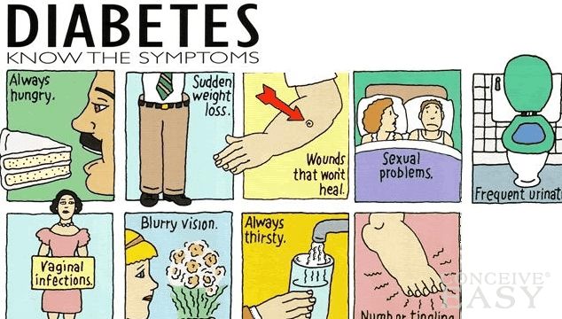
motion of foot), hammertoes, limb length difference, diabetes, bunions and many other conditions.  Orthotics can be custom made 
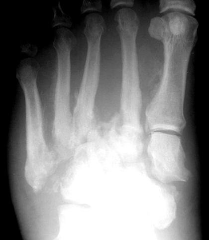
or bought over the counter.  A 3-dimensional scan of the foot is used to send to a lab that makes custom made orthotics, and the orthotics lab can add correction specific to the patients’ needs based on specific instructions in the form of a prescription from our offices.

Orthotics fall into two different categories: functional and accommodative:  

Functional orthotic devices are used to correct biomechanical deformities in the frontal plane, and reduce impact while running or walking.  Functional correction is used to reduce abnormal pronation by providing support of the arch, while accommodative orthotics are a soft supportive device used to provide cushioning, and distribute weight bearing pressures evenly across the bottom of the foot.  When making orthotics it is important to compensate for limb length differences because even a small difference can cause pathology, and affect gait.  Symptomatic differences in limb length usually occur when there is a 4cm or greater difference.  Functional orthotics devices are made of semi-rigid material to provide stability, such as graphite or plastic.  

Accommodative orthotic devices are usually made of softer materials, such as leather or foam to provide comfort.  Accommodative orthotics devices are used in conditions such as diabetes and rheumatoid arthritis to help relieve pressure and apply gentle offloading forces away from prominent structures of the foot. 


This is just a brief overview of the various uses and types of orthotics, and is not complete by any means.  For some people, orthotics devices are a way to treat foot pain conservatively and avoid surgery.  Orthotics devices are sometimes recommended post-operatively to prevent recurrence of various deformities.  However, some patients use orthotics devices to provide stability and support to the foot, and prevent injuries.  

Here, we have a 3-dimensional scanner which can allow us extremely accurate impressions of the feet in order to achieve both optimal comfort and control with our custom molded devices. 


Orthotics, also known as orthoses, refers to any device inserted into a shoe, ranging from felt pads to custom-made shoe inserts that correct an abnormal or irregular, walking pattern. Sometimes called arch supports, orthotics allow people to stand, walk, and run more efficiently and comfortably. While over-the-counter orthotic are available and may help people with mild symptoms, they normally cannot correct the wide range of symptoms that prescription foot orthoses can since they are not custom made to fit an individual's unique foot structure.

Orthotic devices come in many shapes, sizes, and materials and fall into three main categories: those designed to change foot function, those that are primarily protective in nature, and those that combine functional control and protection.
Rigid Orthotics
Rigid orthotic devices are designed to control function and are used primarily for walking or dress shoes. They are often composed of a firm material, such as plastic or carbon fiber. Rigid orthotics are made from a 3-dimensional scan of the foot or feet. Rigid orthotics control motion in the two major foot joints that lie directly below the ankle joint and may improve or eliminate strains, aches, and pains in the legs, thighs, and lower back.

Soft Orthotics
Soft orthotics are generally used to absorb shock, increase balance, and take pressure off uncomfortable or sore spots. They are usually effective for diabetic, arthritic, and deformed feet. Soft orthotics are typically made up of soft, cushioned materials so that they can be worn against the sole of the foot, extending from the heel past the ball of the foot, including the toes. Like rigid orthotics, soft orthotics are also made from a 3-dimensional scan of the foot.
Semi-Rigid Orthotics
Semi-rigid orthotics provide foot balance for walking or participating in sports. The typical semi-rigid orthotic is made up of layers of soft material, reinforced with more rigid materials. Semi-rigid orthotics are often prescribed for children to treat flatfoot and 
in-toeing or out-toeing disorders. These orthotics are also used to help athletes mitigate pain while they train and compete.

If there is any question whether these sorts of devices may help you, most likely than can, because if you are questioning if you need them most likely your feet have some symptomatic concern which does require an evaluation.


Brian Timm, DPM, FACFAS

Board Certified by the American Board of Podiatric Surgery