Showing posts with label ulcer. Show all posts

Thursday, October 30, 2014

Diabetics who smoke cigarettes beware


Smoking releases a molecule known as norepinephrine, which triggers blood vessels to constrict, or narrow. When the already small vessels in the foot constrict, oxygen and the cells that fight infection and aid in healing are unable to reach tissues. Each cigarette smoked has been shown to cause oxygen levels to fall and remain low for 30 to 50 minutes. This is of great concern in those with a foot wound or incision after surgery, in which blood flow and oxygen are vital to skin healing.    
Each cigarette contains more than 4,000 natural and synthetic chemicals. Some of these chemicals include carbon monoxide, methanol (which is found in rocket fuel), ammonia, and nicotine.  Nicotine has been shown to decrease the molecules that are responsible for growth of new blood vessels and development of osteoblasts, or bone building cells. Carbon monoxide further contributes to the decrease in tissue oxygen caused by vessel constriction, by taking oxygen away from the molecule it is normally transported in in the blood. It is for these reasons that quitting smoking is imperative for individuals with foot problems. Studies have shown that there is a 2.7 times higher risk of bone not healing in smokers compared to non-smokers undergoing foot surgery.  

Prior to foot surgery involving bone, smoking should be stopped at least 6 weeks prior to the procedure if possible and then take that opportunity to stop smoking indefinitely


In individuals with diabetes or other disorders affecting their blood vessels, smoking is even more dangerous.  Pressure ulcers develop when sensation is diminished and the normal feeling of pain associated with a wound is not present. Tissue healing is impaired and there is a heightened risk of infection developing. Combined with smoking, such risks are furthered increased. Individuals with diabetes who notice a foot wound, whether they are smokers or not, should contact their podiatrist immediately. Early treatment can prevent infection and worsening of the wound that can potentially lead to a need for amputation.  Bill’s story in the smoking ad is a scary one, but by sharing it he may be able to prevent others from suffering a similar fate of amputation. 


Tuesday, September 16, 2014

CROW Brace to treat severe deformity in high risk patients.

CROW - Charcot Restraint Orthotic Walker

What is a CROW Brace?


The Charcot Restraint Orthotic Walker, or CROW, is a stable boot designed to accommodate and support a foot with Charcot
neuroarthropathy. The CROW consists of a fully enclosed ankle/foot orthotic with a rocker-bottom sole.  It is a common treatment used after the acute charcot foot has calmed down.
 

What is Charcot deformity?

This occurs when bones and joints in the foot fracture, break up or pop out of place with minimal or no known direct injury. In the United States, this deformity is most commonly seen in people with diabetes. The foot first enters an acute stage of swelling, warmth and redness, which eventually diminish. Broken bones and dislocations can occur, causing severe deformities of the foot and ankle. Some patients develop pain or ulcers when the affected foot becomes deformed. CN can affect the other foot or happen again in the same foot. The foot does not regain its normal shape. 

What is a CROW made of?

The boot is custom made for each patient’s foot. The outer shell consists of two plastic or fiberglass clamshell pieces that fit and are strapped together with Velcro. It is sturdy and can prevent other bones from cracking or breaking and can be walked on. The bottom of the boot has a rounded rocker-bottom shape. The boot contains a custom, removable foam insole. Each insole is adjusted to distribute weight equally and also to support the ankle joint.
 

What does the boot do?

The CROW functions by providing even support to the entire foot, especially to areas that are overstressed due to the neuroarthropathy. These deformities often cause the foot to bend out of shape. The resulting stress on the foot can cause ulcers, which can develop into severe infections if left untreated. By distributing pressure equally throughout the leg and foot, the CROW removes excessive forces and gives the foot time to heal. It is easier to use than a cast, can be removed for wound care and washing, and is more durable.
 

Which patients can use the CROW?

Patients with acute Charcot can begin using the CROW after the swelling has receded. This can take months.  Patients with mild to moderate deformities will benefit most from the CROW. Patients with severe  deformities or extreme foot/ankle instability may need surgery instead of using the CROW.
 

How does it affect daily life?

Fortunately, the CROW is adaptable to daily life. Because of the clamshell design, the patient can easily remove the boot in order to keep the foot clean and sleep better. In addition, its fitted shape and good support allow people to return to walking, standing and driving more normally.
 

What are typical outcomes?

The most important outcome is that patients are able to continue to bear weight while minimizing pressure and giving the foot a chance to heal. Healing may require many months. However, the disease process may return and/or affect the other foot, so regular and lifelong monitoring of the condition is necessary.
 

What are the possible complications?

Despite the sturdiness of the boot and the distribution of forces, the bones of the foot could still break. The foot could develop open sores, though the boot is designed to prevent it. As always with Charcot deformity, some joints may heal incorrectly or not at all. Unfortunately, other factors such as poor glucose control and bad nutrition can prevent healing despite use of a CROW.
 

Frequently Asked Questions

What options do I have when my foot is still swollen?Patients often wear special casts until their feet stop swelling enough for them to use a CROW. The cast serves to stabilize the foot and prevent unstable motion, similar to the CROW. However, unlike the CROW, these casts cannot be removed.

Monday, September 8, 2014

Limb Preservation: Total Contact Casting


Diabetics with neuropathic ulcers affect at least one quarter of all diabetics in the United States, and according to Wounds Research, approximately 1 in 5 of these individuals will eventually require an amputation of their foot. Total contact casting has recently become more popular as an option for foot protection, and to mediate the excessive wear and tear that some diabetic foot sores suffer.

What is total contact casting?

As the name suggests, this technique, which is used by many diabetic foot specialists, is a thin cast that is applied to the entire plantar surface area of the foot, as well as the lower leg. Based on its design, the cast can take up as much at 92 percent of the pressure from the ulcer site. Since ulcers often occur on parts of the foot or leg that are repeatedly impacted through daily movement, it is important that they receive as much padding as possible without affecting an individual’s normal walking patterns. Although total contact casting provides relief from certain pressures, the cast works with the natural human gait to make sure that no section of the foot or leg is required to take on excessive weight or pressure.

Does it always work?

Total contact casting has been shown to be effective in the healing of foot and lower leg ulcers, but only those that are noninfected. According to current research, the success rate is between 72 and 100 percent in most patients. A study conducted at the University Hospital Maastricht in the Netherlands examined how total contact casts would fare on infected patients, and of the 28 patients in the study group, only 36 percent showed signs of healing. The study group recommended that individuals with infected diabetic ulcers look toward alternative treatments for healing, according to the American Diabetes Association.

Surgery may become necessary in conjunction with acute infections.


Monday, March 3, 2014

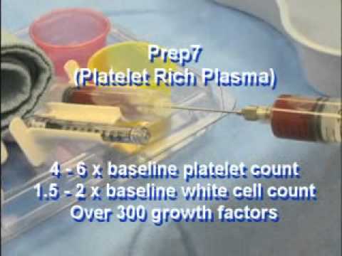
PRP Literature Review


As the use of PRP increases, additional studies may establish PRP as an efficacious treatment modality and guide future treatment of chronic diabetic foot ulceration.

- Yale Journal of Biology and Medicine March 2010


We have been using PRP injections in this manner since 2010 as well, and this study was published promptly (and independently) from our center, but it shows the progressive mindset and improved outcomes from our center.

We are always striving to obtain the latest in technology to help treat our patients both effectively and not only by use of standard of care methods we will often go ABOVE the standard of care to ensure the most optimal in outcomes for our patients with diabetic wounds and neuropathic ulcerations.


McAleer et al. (2006) found that the use of autologous PRP was successful in healing a chronic lower extremity wound in a case study of a 57-year-old man with type 2 diabetes and a wound of six months duration. 


Salemi et al. (2008) was a more recent case study evaluating the effectiveness of a combination of autologous adipose tissue and PRP in a lower extremity ulcer of three years duration in a non-diabetic 65-year-old male patient.

Margolis et al. (2001) was a retrospective cohort study devised to estimate the effectiveness of platelet releasate (PR) in the treatment of diabetic neuropathic foot ulcers. Of the 26,599 patients included in the study, 21 percent were treated with PR by the end of the 12-week run-in period before the 20-week study period began.  The investigators concluded that PR was more likely to be used in more severe wounds and was also more effective in treating these wounds than the standard of care. 

Driver et al. (2006) carried out the first reported prospective, randomized, controlled multicenter trial in the United States regarding the use of autologous PRP for the treatment of diabetic foot ulcers. Participants included 72 patients with type 1 and type 2 diabetes between the ages of 18 and 95 from 14 investigation sites suffering from an ulcer of at least four weeks duration. In this study, investigators compared the effectiveness of autologous PRP gel to that of normal saline gel for 12 weeks. The primary objective of this study was to evaluate the safety of PRP and the incidence of complete wound closure, defined as 100 percent re-epithelialization, when compared to the control treatment, and a secondary objective was rate of wound closure. Patients were randomized into two groups — standard of care with PRP gel or control (saline gel) — and were evaluated biweekly for 12 weeks. After excluding 32 patients from the final per-protocol analysis because of failure to complete treatment and protocol violations, the authors found that 68.4 percent (13/19) of patients in the PRP group and 42.9 percent (9/21) in the control group had wounds that healed. Wounds in the PRP group healed after a mean of 42.9 days (SD 18.3) vs. 47.4 days (SD 22.0) in the control group. 

This is significant, because the longer the duration of time that there is an ulceration on the plantar skin the more directly proportional to likelihood of osteomyelitis formation occurs. 


Metalink: http://www.ncbi.nlm.nih.gov/pmc/articles/PMC2844688/