Thursday, February 12, 2015

5 Facts about Charcot Neuroarthropathy you need to know


1) With an unknown definitive mechanism of incidence, this disease process has many proposed hypothetical mechanisms currently in clinical practice. Some believe that increased inflammation and microfracturing of the bone secondary to sensory loss are key components, others believe glucose levels and specific traumatic events are responsible for this process which are compounded by inability to offload and protect the injured limb leading to progressive breakdown of the bone. We all agree that these elements are part of the problem, but do not necessarily occur in all cases. Especially in non diabetics with the disease of Charcot neuroarthropathy.

2) We know that many cases start with increased warmth, redness, and swelling. Unfortunately not ALL cases give us this "textbook" presentation.

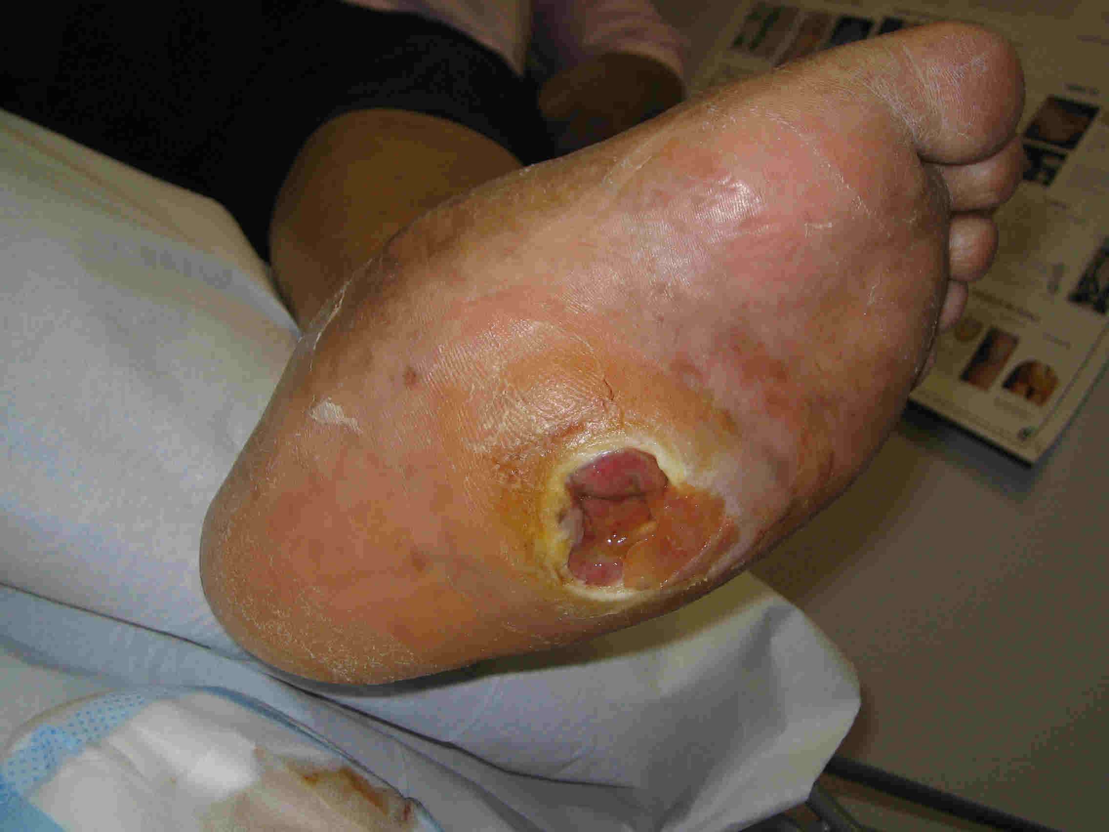
3) We also know that characteristic radiographic changes may occur.  And when they do, differing mindsets and clinical approachs are valid and are practiced routinely in my practice. If an ulceration is present, or progressive radiographic changes are found, surgery is likely required for limb salvage.

4) We generally understand that patients with low or no protective sensory threshold on their feet tend to have a higher incidence of this process.  Also patients that have poor glucose control and are obese tend to have this same increase in incidence. Edema and general leg swelling in diabetic patients with Hemoglobin A1c levels higher than 8.0 tend to have increased risk of Charcot development in their feet. Usually all of these patients have good blood flow with regards to arterial circulation, but may have venous or lymphatic flow compromise.


5) Not all patients with Charcot neuroarthropathy are diabetic. I have treated many patients whom are not diagnosed with diabetes, and did not present with red swollen foot, who developed Charcot neuroarthopathy with radiographic bone degradation and resultant foot deformity. These patients do tend to have profound sensory polyneuropathy of unknown etiology (idiopathic) and are still at risk given the above listed requirements. I have had to reconstruct several patient's feet without diabetes as an underlying diagnosis, and it seems that they tend to have less overall complications but are still prone to the neuroarthropathy nonetheless.


The take away from this blog is that many of the "facts" about this disease process have some "grey area" information. You should seek the expert opinion with good experience in this disease process prior to any surgical intervention or decision for lower leg or foot amputation. The doctors at FFLC are well equipped to accommodate and treat this condition and are well versed in all avenues regarding limb salvage.

Monday, January 26, 2015

Milan Defender Zapata Sidelined Due to Foot Injury


Serie A club Milan defender Cristian Zapata suffers from a fractured foot during the final minutes against Sassuolo.

“AC Milan communicates that in the final minutes of Milan-Sassuolo, Cristian Zapata, following a challenge with another player, suffered a bone fracture at the base of the second metatarsal in his right foot.”

Metatarsal fractures are common injuries of the foot, typical caused by direct crushing injury or by twisting injury where the forefoot is fixed and hindfoot or leg rotating (more common, associated with sports injury). The 5rd metatarsal bone is most commonly fractured metatarsal bone, while the 3rd rarely gets fractured in isolation. A British journalist named the 2nd metatarsal bone the “Beckham bone” due to a fracture David Beckham got after he was tackled by Aldo Duscher, this received a lot of publicity during that time.


Generally these injuries may be treated nonsurgically with a walking boot, or cast. Irregardless of treatment, it is likely this injury will sideline this footballer for a minimum of 8 weeks.  

Wednesday, January 7, 2015

New Year's Resolutions - Walking and Osteoporosis


Start walking today, as this is a very good way to increase bone density. Osteoporosis is the most common skeletal disorder.  It is characterized by an absolute reduction in bone density and mass.  Bones become weak and are at increased risk of breaking.  Because 28 of the bones in the body are located in the foot and they are under the pressure of the entire body weight, fractures commonly manifest here.  Sunlight does not directly provide vitamin D; it stimulates the body to make vitamin D from a precursor found in the skin.  The active form of vitamin D promotes the uptake of calcium from the intestines so it can be used to calcify bone as well as stimulating the parathyroid hormone to reabsorb calcium before it is filtered out by the kidneys as waste.  Without enough vitamin D, not only are individuals at risk for osteoporosis, but they can also develop a similar condition called osteomalacia.  In osteomalacia, bone cannot be mineralized and symptoms include muscle weakness, bone pain and bone deformities.    The childhood form of osteomalacia that is more commonly known is called rickets.  In rickets, children affected will typically be six to 12 months of age and will have symptoms of muscle tetany or extreme muscle contraction, delayed development, smaller overall stature and soft tissue swelling, or edema around the growth plates located at the ends of bones.  Bowing of the tibia bone inward in the legs is a common consequence of vitamin D deficiency in rickets. 
The one upside of these diseases is that there are some preventative measures that can be taken to avoid their occurrence.  To keep your foot bones and the rest of the bones in your body healthy, be sure you have enough vitamin D as a part of your healthy diet.  Bone mass peaks at age 35, so it is especially important to maintain an adequate amount of vitamin D and calcium in your diet and through exposure to sunlight later in life.  Females are prone to developing osteoporosis following menopause and therefore should also take extra care to keep their bones strong.  By contacting your podiatrist at the first signs of foot injury or bony pain in your feet and ankles, both any fractures that have occurred as well as loss of bone density can often be identified in one set of x-rays.    The sooner that loss of bone density is identified, the sooner steps can be taken to supplement vitamins and minerals necessary to keep you healthy and on your feet!