Showing posts with label ankle. Show all posts

Thursday, December 11, 2014

Holiday Foot Care Reminders


With the holidays fast approaching, many individuals may feel the mounting pressure of finding the perfect gift by spending hours at the mall.  The pressure placed on feet dashing from store to store can add up, and in individuals with diabetes can become dangerous.   Even though the holiday of St. Nicholas passed this past week, in which children leave their shoes out to be filled with treats, individuals with diabetes may still want to leave their old shoes out for good and opt for a better, pressure relieving pair.  

High pressure in a healthy foot signals pain, and subsequent alleviation of the inciting pressure by the person moving their foot or changing their shoe to stop the pain from occurring.  In diabetic individuals with peripheral neuropathy, pain and areas of high pressure cannot be felt due to loss of sensation.  This loss of sensation causes of loss of protection to the foot and wounds may develop in areas of abnormally high pressure.  Any deformity that predisposes the foot to rub in a shoe creates an area that is more likely to ulcerate.  Surgery to remove deformities including bunions, hammer toes and bony bumps in the ball of the foot are one of the ways that your podiatrist may remove an area of increased pressure that is likely to develop or has previously developed an ulcer.  Surgery to lengthen the tendon that attaches the calf muscles to the heel bone is also used to lessen pressure on the ball of the foot.  When this tendon is too tight, the heel is lifted and the front of the foot is levered down towards the ground, creating an area of high pressure. 


Along with surgery, regular callus and corn removal by your podiatrist is also very helpful in removing pressure.  Shoes, casting and custom orthotics are all used to better disperse pressure on the foot.  These treatment means are also useful to prevent ulcers from developing or recurring.  By custom molding shoes to the foot, deformities are accommodated for and areas of high pressure are relieved.  Custom orthotics for diabetic patients with neuropathy are made with several layers of material so that any friction that would normally be imparted to the foot is instead absorbed by the orthotic.  Talk to your podiatrist about taking pressure off this holiday season and keeping your feet healthy!


Brian Timm, DPM, FACFAS

Thursday, November 20, 2014

External fixation here in Naples Florida


External fixation may look very high-tech, and maybe even scary.  However, external fixation has been used in one way or another since almost 2400 years ago.  External fixation techniques were described by Hippocrates, and were used in treating tibia fractures.  External fixation is a minimally invasive technique to reduce displaced fractures and has become a very important part of deformity correction and nonunion and pseudo arthrosis repair as well.

Jean François Malgaigne was one of the many pioneers that made advancements with external fixation devices.  In 1846, Malgaigne used a device that consisted of a clamp and four metal prongs to reduce and stabilize a fracture of the patella, or kneecap.  Following this external fixation device many other similar inventions were used to treat fractures in various locations.  In 1938, Raoul Hoffman made advancements that made external fixation even more useful, and allowed surgeons to place pins into a fracture for stabilization with guidance, while being minimally invasive.  In 1951, Dr. Gavriil A. Ilizarov developed the external fixation device that is still in use today.  Ilizarov’s fixation device consists of a metal frame that encircles the limb, and is attached to underlying bone by pins.  Threaded rods and hinges allow movement of the bone to the correct alignment.  Ilizarov’s external fixator is great because adjustments can be made without opening the fracture site, and the device provides stability. Also it can be converted to internal fixation once the soft tissues have become less swollen. 


Modern day external fixation not only provides stability to a fracture, but can also be used for soft tissue deformities, as well as other bony deformities.  External fixation is preferred when slow correction is required, and even more useful in high risk patients with vascular disease and even open fractures with high risk of infection.  The chance of getting a blood clot is lessened because with external fixation patients can be partial weight bearing, or weight bearing as tolerated following the procedure due to the stability that the fixation provides.  External fixators have been used for other bony deformities, such as Charcot, ankle arthritis, and clubfoot.  External fixation can also be used to lengthen amputated foot and toe stumps.  External fixation has definitely helped many people, but there are still some cons.  Pain and infections are two issues associated with external fixation, and rates vary depending on the extent of the procedure, and the location of the device.  External fixation devices are used by specially trained physicians and all three of the doctors at FFLC are capable of utilizing these various techniques for patients that require such interventions.  


Brian Timm, DPM, FACFAS
Board Certified by the American Board of Foot and Ankle Surgery in Foot and Reconstructive Rearfoot and Ankle surgery

Monday, November 10, 2014

Swimming and your feet


Everyone is starting to return to Florida for the seasonal upgrade in weather.  Fire up the pool warmer, and get ready to swim.

 Swimming is known for being an activity that is less damaging to the joints of the knee, foot and ankle, and can even offer a means of exercise for athletes of other sports with certain foot injuries!
Just about the worst thing a runner can hear is that they are injured and going to have to take time off from running.  Luckily, swimming can help soften this blow by offering an alternative way to get some cardiovascular activity.  Stress fractures are small fracture that occurs from excessive force on normal bone and can force runners to talk a break from running for several weeks.  For the runner with a stress fracture, swimming is often a great alternative that will not bear weight on the injury.  Swimming can also help an athlete ease back into their favorite sport after other serious injuries.  For example, after a peroneal tendon tear, swimming can help to make the transition from being immobilized in a cast or a CAM walker boot to normal running or other weight bearing sports go more gently by allowing the muscles to slowly adapt.  
Swimmers should also be careful to take care to avoid injuries that can occur from intense pool training.  Ankle pain is a common complaint from the repetitive motion the ankles are forced through while swimming.  The Achilles tendon, which is responsible for flexing the foot, may also become irritated from the repetition and develop Achilles tendonitis.  While the Achilles is doing much of the work, the extensor tendons on the top of the foot can also become irritated by being tightly pulled.  By taking care to stretch the feet and ankles properly and always listen to your body when working out, these injuries can be minimized.  Swimmers also need to take extra care to avoid developing warts, Athlete’s foot, and fungal nails from bacteria and fungus that may live in locker rooms and poolside surfaces where other bare feet have deposited them.  If these conditions do develop, see us at Family Foot and Leg Center for an urgent evaluation, as to help prevent spread to others. 

Dr Timm, DPM, FACFAS

Board Certified by the American Board of Foot and Ankle Surgery in Foot and Reconstructive Rearfoot and Ankle Surgery.


Wednesday, September 3, 2014

Overview of the Charcot disease


What is it?

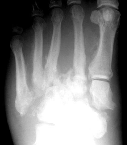
Charcot arthropathy, also known as Charcot foot and ankle, is a syndrome in patients who have neuropathy or loss of sensation. It includes fractures and dislocations of bones and joints that occur with minimal or no known trauma.​

Symptoms and Clinical Presentation

​Initially, there may be swelling, redness and increased warmth of the foot and ankle. Later, when fractures and dislocations occur, there may be severe deformities of the foot and ankle, including collapse of the midfoot arch (often called rocker bottom foot) or instability of the ankle and hindfoot. The syndrome progresses through three general stages:

•
Stage 1 (acute, development-fragmentation): marked redness, swelling, warmth; early radiographs show soft tissue swelling, and bony fragmentation and joint dislocation may be noted several weeks after onset
•
Stage 2 (subacute, coalescence): decreased redness, swelling and warmth; radiographs show early bony healing
•
Stage 3 (chronic, reconstruction-consolidation): redness, swelling, warmth resolved; bony healing or nonunion and residual deformity are frequently present.

Cause (including risk factors) 

Charcot foot occurs in patients with peripheral neuropathy resulting from diverse conditions including diabetes mellitus, leprosy, syphilis, poliomyelitis, chronic alcoholism or syringomyelia. Repetitive microtrauma that exceeds the rate of healing may cause fractures and dislocations. Changes in circulation may cause resorption of bone, weakening the bone and increasing susceptibility to fracture and dislocation. 

Anatomy 

Charcot arthropathy may affect any part of the foot and ankle, including (in decreasing order of frequency) the midfoot, hindfoot, ankle, heel and forefoot.  Multiple regions may be involved concurrently.  Fractures and dislocations frequently involve several bones and joints, with extensive fragmentation and deformity. 

Diagnosis 

Time between onset of symptoms and diagnosis may be several weeks or months. Delay in diagnosis may or may not affect the end result because gross instability may occur even if prompt diagnosis is made. Diagnosis is based on a high index of suspicion for this problem in patients with neuropathy. Increased redness, swelling and warmth may be the only early signs. Some patients have pain. Early radiographs may show soft tissue swelling with no bony changes, but repeat radiographs several weeks later may show bone and joint changes.

Treatment Options 


Non-Surgical: Non-operative treatment includes a protective splint, walking brace, orthosis or cast. Early weightbearing is allowed in stage 1 by 41 percent of specialists and in stage 2 by 49 percent of specialists, and other specialists recommend non-weightbearing. After stable healing is noted in stage 3, treatment includes accommodative footwear with protective orthoses.Surgical: Selected patients with instability in the early stages may be treated with open reduction and internal fixation and fusion. In the later stages, surgical options may include realignment osteotomy and fusion (correction of deformity) or ostectomy (removal of bony prominence that could cause an ulcer ). 


* Sometimes the surgery can fail, and this is commonly because of improper or inadequate fixation, poor glucose management, obesity, noncompliance, and even infection. 

Wednesday, August 27, 2014

Charcot and you.


Many diabetic patients may have heard warnings to look out for “charcot foot” but they may not know exactly what this means.  Different from diabetic charcot foot is an inherited disease with another characteristic foot type called Charcot-Marie-Tooth disease.  As a part of September’s Charcot-Marie-Tooth (CMT) disease awareness month, it is critical for patients to understand the differences between these two neurological disorders. 

CMT
Charcot-Marie-Tooth disease is caused by a gene defect that is often inherited.  If CMT is seen in other family members, parents should be on the lookout for the development of slowly progressing muscle weakness in the lower extremities before age 20.  Individuals with CMT have nerves that lose their myelin covering, which normally allows signals to be sent to and from skin and muscles at a normal speed allowing sensation and muscle contraction.  Patients will not usually complain of any numbness because their sensation was likely never completely normal.  As a result of the weakness and loss of sensation, first in the legs then typically seen in the upper extremities, kids with CMT may seem clumsy and have difficulty walking without tripping or rolling their ankles. The feet will often have a high arch appearance and may be prone to ulcers from damage caused by lack of ability to sense pain. 

Diabetic Charcot Foot
Patients with diabetes need to be concerned about charcot arthropathy when they develop areas of the foot where they can no longer feel anything.  The combination of lack of sensation allowing damage to the foot that patient cannot feel and an increased blood flow supplying mediators of the inflammatory process allows a “charcot joint” to develop.  In the charcot joint, the repeated small injuries occurring cause bones to gradually fracture and dislocate.   This causes deformity (often an extreme flat foot) which typically places the diabetic patient’s foot at a greatly increased risk of developing ulcers and subsequent infection with severe consequences.  The most common signs of the development of charcot arthropathy are swelling and increased temperature of the affected area of the foot, redness, pain and the feeling of a “loose bag of bones” when the joint is moved. 

In both diabetes with loss of sensation, and Charcot-Marie-Tooth, foot self-exams are immeasurably important in preventing ulceration and further complications.  Protective shoe gear and custom made orthotics can provide additional protective measures for the foot.  As shown by these two disease processes, loss of sensation in the foot is a major problem that should always be evaluated by your foot and ankle specialists here in Naples Florida.  

Monday, June 9, 2014

6 Secrets to Manage Foot Arthritis


Arthritis is the leading cause of disability in the United States.  Almost half of people in their sixties and seventies suffer from arthritic foot pain.  There is no cure for arthritis, but there are many types of treatments available that focus on pain management.  Osteoarthritis typically occurs in older individuals, or people that have had a previous injury.  Osteoarthritis is a degenerative joint disease. Over time the cartilage between joints wears down leading to inflammation, swelling, and pain.  In younger individuals that develop osteoarthritis there is usually a history of previous injury, family history of osteoarthritis, or obesity.  

The foot consists of 33 joints, but there are a select few that are more prone to osteoarthritis than others.  The ankle, the great toe, 3 joints in the hindfoot (talocalcaneal, calcaneocuboid, talonavicular joints), and the midfoot (metatarsocuneiform joint) are the most commonly affected joints.  Osteoarthritis can also be caused by abnormal foot structure, such as flat foot or a high arch.
Some people believe that cold, damp weather exacerbates their osteoarthritis, and some even say they can predict the weather.  Common symptoms of osteoarthritis include: stiffness, limitation of the joint, swelling, difficulty walking and tenderness that is persistent.  Osteoarthritis is a chronic condition, and can be debilitating.

There are many great treatments available, but here are just a few solutions for osteoarthritic foot pain that you can try prior to seeing us at the Family Foot and Leg Center:

1.      See me at least once a year to monitor changes, and modify treatment
2.      Comfortable and supportive shoes are necessary, and sometimes orthotics or ankle braces are also helpful
3.      Weight loss
4.      Anti-inflammatory medications such as NSAIDS (ibuprofen) or corticosteroid injections that can be given by your podiatrist
5.      Physical therapy and exercises
6.      Try a topical medication that has capsaicin, an ingredient that can help reduce pain

Dr Brian Timm
Board Certified by American Board of Podiatric Surgery


Monday, June 2, 2014

Masses on your foot


There are many types of tumors that affect the foot, and many are benign.    

Two of the most common types are ganglion cysts and plantar fibromas.  

Ganglion cysts are also known as bible cysts because common treatment was hitting them with a bible so that the cyst would burst.  Ganglion cysts are more common in younger people, while plantar fibromas are more common in older people.  
Ganglion cysts often occur on the top of the foot, but are also very common on the hand.  The cysts are fluid-filled, occur at joints, and often change size. Ganglion cysts are usually observed for any changes unless they reach a size that causes pain or pressure on nerves or arteries.   Immobilization of the area is also sometimes helpful because activity often causes the cyst to increase in size.  If they reach a large size, then further treatment may be beneficial.  Further treatment of ganglion cysts usually consists of draining the fluid with a needle, or aspiration.  Aspiration of the fluid is not always successful because the root of the cyst may remain, and the cyst will return.  Surgical treatment may be recommended for recurring ganglion cysts. 


Plantar fibromatosis is the most common benign tumor on the bottom of the foot.  Plantar fibromatosis is also known as Ledderhose’s disease, which is a thickening of the foot’s plantar fascia.  Over time the lump increases in size, and may start causing pain or limitations in activity.  This type of benign tumor is often associated with a family history of the disease, or patients with diabetes.  Initial treatment is off-loading to avoid direct pressure or worsening of the tumor.  Corticosteroid injections and cryotherapy are often used initially before surgical options are considered.  Surgical removal of a plantar fibroma is more difficult than removing a ganglion cyst, which is why more conservative therapy is recommended unless conservative methods have failed. 
Ganglion cysts and plantar fibromatosis are only two of many types of possible tumors, but they are the most common benign tumors presenting in the foot.  These are both very manageable conditions, but with any lump or bump in the foot it is important to see your Podiatrist to rule out something more serious.

Brian Timm, DPM, FACFAS
Diplomate: American Board of Podiatric Surgery
Board Certified in Foot Surgery

Tuesday, April 29, 2014

Rollerblading and your ankle health.


Although rollerblading is a low-impact sport, The Consumer Product Safety Commission reported an average of over 32,000 inline skating injuries every year from 2003 to 2007, according to the National Safety Council. Most injuries affect the upper body, but ankle injuries are one of the most common injuries to the lower extremities.The firm boot of an inline skate provides good ankle support when the skate fits properly, but fit is key. Protecting your ankles means not just following proper skating technique but also shopping smartly for your inline skates.

Often times the ankle will be supported well within the confines of the device. However knee sprains and syndesmotic injury can occur (high ankle sprain) in the right setting while rollerblading. More often foot injury is likely, with blisters, metatarsalgia, and even mild neuropraxia in the wrong rollerblade configuration. This is a general guide to choose the right blades for your feet.

Dr Timm
Diplomate American Board of Podiatric Surgery
Step 1
Shop for skates after a day of activity to compensate for any swelling of the feet that occur while rollerblading.
Step 2
Pick skates for your gender, especially if you are a woman. While women can wear unisex skates, those designed especially for a woman better fit a woman’s narrower Achilles tendon and lower position of the calf muscle, notes Liz Miller, author of “Beginner's Guide to Inline Skating.”
Step 3
Look for skates that lace up and buckle at the top. The combination provides a more secure fit for ankle support.
Step 4
Squeeze the plastic support in the boot. It should be firm and unmovable.
Step 5
Try on skates wearing a pair of thick, absorbent athletic socks -- the same type of socks you’ll wear when skating.
Step 6
Wear the skates for at least 15 minutes before making a purchase. When laced and buckled, the skates should feel snug, but not so tight that any part of your foot feels pressure.

Skating

Step 1
Stretch before skating. To prevent ankle injuries, focus on your feet: Flex and point your toes and rotate your ankles in circles.
Step 2
Kick your feet into the back of your rollerblades before lacing and buckling up.
Step 3
Warm up by gently skating around for five minutes.
Step 4
Match your skating location to your skating ability. If you are a new skater, begin on a large patch of level grass. Empty tennis courts and parking lots are also good places to learn.
Step 5
Bend your knees when you skate to lower your center of gravity. Skate with a wide stance if you are new to the sport. Skate with a shoulder-width stance if you are an experienced skater. A narrower stance gives you better control for tricks, but a wide stance helps beginners maintain balance.

Tuesday, April 15, 2014

Credentials you can trust.

Often times patients will ask me what my credentials actually mean. Below is a summary directly from the website of the American Board of Podiatric Surgery (which will change the name to American Board of Foot and Ankle Surgery this year) which delineates the prerequisites necessary to be deemed "Board Certified" by this particular surgical board. 

-Dr Timm

Diplomate American Board of Podiatric Surgery

Certification in Foot Surgery

Certification in Foot Surgery              indicates that a podiatrist has demonstrated a cognitive knowledge of podiatric surgery, including the diagnosis of general medical problems and surgical management of foot diseases, deformities, and/or trauma, and those structures which affect the foot and ankle.                                                       THE CURRENT QUALIFICATIONS INCLUDE:                                           

Completion of a four-year doctoral training at a CPME-accredited podiatric medical school.                         

Completion of residency training program approved by the Council on Podiatric Medical Education (CPME).                                                                                                                                                                            

Submission of a list of procedures through Podiatry Logging Service for Surgery demonstrating a diversity of surgical experience in foot surgery.    

Approval by the ABPS Credentials Committee of complete foot surgery case documentation of procedures selected by ABPS from the list of procedures.       

Passing Part I and Part II of the Certification in Foot Surgery Examination.Diplomates certified in Foot Surgery must recertify every ten years.