Tuesday, March 18, 2014
Secrets of Chronic Achilles Pain
1) NOT all posterior heel pains are tendon problems.
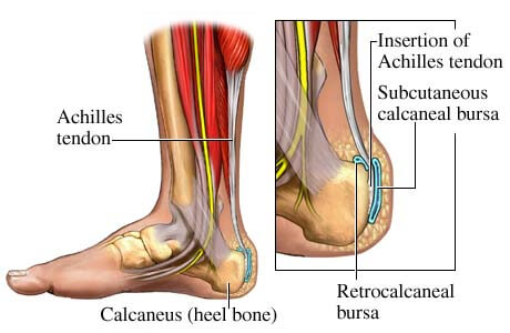
We will provide a thorough examination, which includes diagnostic ultrasound, radio graphs, and clinical exam with history taking to provide the best possible outcome for your pain. There are several other structures which are present and could be treated in a different manner than any straight forward Achilles tendinitis. Noted above, there is a bursa which is present here and can often be the root cause of pain secondary to bursitis. This is treated differently than Achilles tendinitis. This may also require MRI to determine how much of the inflammation is related to bone versus soft tissue. Additionally, and more rarely, a stress fracture can cause this pain in the calcaneus. This is not common but we check for this as well.
2) Tendon tears do NOT always require surgery.
After clinical examination and proper staging/grading of the extent of the tear, we have had success in treating Achilles partial tears and even ruptures non-surgically in select patient populations. We have a variety of techniques at our disposal in order to facilitate the healing process if surgery is not indicated. In the case of chronic tears there are several nonsurgical treatments that work, particularly PRP injections, High energy shock-wave treatments, and even physical therapy with anti inflammatory ultrasound.
3) Bone spurs are NOT always relevant.
Often times patients will state that they "Have had a spur in the past but it went away". Usually any bone protuberance which is either palpable or visible on a plan radio-graph or MRI will NOT go away spontaneously ever and always if indicated will need resection if it is the root cause of the symptoms. In most cases the spur is not the root cause of the symptoms, and is not required to remove it, but there are far too often very large spurs which are not even symptomatic that are visible on X-ray examinations in many of our patients, therefore backing up further the concept that bone spurs are not always the real problem.
Bio-mechanically a bone spur will exist in areas of either repeated chronic trauma, or areas of chronic taut insertions. Rarely is this an isolated tumor of bone, and that being said, it is often not necessary to remove the bone spur.
Dr Timm
Board Certified by the American Board of Podiatric Surgery
Labels:
Achilles,
Bone spurs,
Heel pain,
Heel surgery,
injury,
MRI,
pain,
PRP,
Shock wave,
surgery,
tears,
tendinitis,
treatment
Monday, March 10, 2014
Secrets of Recurring Heel Pain
Many patients are becoming even more active with age, and as this new generation of elderly patients become our community's senior citizens we are finding out more often that you are only as young as you feel. With this, there are different aches and pains, and they are more common and can be restricting towards maintaining this level of leisure. Above all other pains common to the foot and ankle, heel pain is by far the most common. Sometimes, we will be a 3rd and 4th opinion to attempt to remove this nuisance from our patient's feet. Most often heel pain is treated by cortisone injections, physical therapy, weight loss, and orthotics.
But what about the patients who have been there and done that?
There are few treatments that are more promising in the field of heel pain management with more potential than the
Extra Corporal Shockwave Therapy for very tough heel pain. A good overview of the proposed mechanism for how this works involves using devices that generate pulses of high-pressure sound that travel through the skin and yeild natural repair from the tissues and increased blood flow networks to damaged tissues.
For reasons that are not fully understood, soft tissue and bone that are subjected to these pulses of high-pressure energy heal back stronger and without causing further damage to the tissues like repeated cortisone injections may do.
The FDA has approved the use of some ESWT machines for heel pain and tennis elbow. ESWT devices evolved from lithotripters (a.k.a. kidney stone shock wave machines). The discovery of the beneficial effects of ESWT came as German researchers were trying to determine what type of high-pressure pulses could be sent through the body to disintegrate kidney stones without causing harm to surrounding tissue. In laboratory animals and humans, it was discovered (with some surprise) that surrounding tissue would often heal back stronger and this applies well to our topic of heel pain.
Physical therapists use ultrasound machines that warm internal tissue by high frequency sound waves, but the ESWT machines send higher-energy pulses 2 or 3 times per second rather than continuous lower-energy waves. Electricity is not sent into the body. It may take as long as 5 months to see the full benefit of an ESWT treatment. The beneficial effect of the high-pressure waves may be from the growth of new blood vessels ("neovascularization") in small cavities that are created by the pulses. New blood vessels to an area of tissue would promote healing either directly or indirectly by providing additional growth factors to the area of concern by way of new vascular channels.
Some studies have even shown this therapy to be equivalent to a fasciotomy, which is an invasive procedure that involves an incision to sever the medial fibers of the fascia for reduced pressure and tension on the inferior heel bone. This means that you can get equivalent results without the complications of a minor procedure. We provide 2 variations of the ESWT, a high energy and a lower energy, and each has benefits that have been shown to work well on difficult heel pain. Either method will be beneficial to our patients, and if you are limping in the AM, or have trouble walking after a brief rest from activity because your heel is holding you back, you should consider a consultation with one of our doctors.
Dr Timm
Diplomate: American Board of Podiatric Surgery
Fellow: American College of Foot and Ankle Surgery
Labels:
Board certified,
ESWT,
FACFAS,
FDA,
Heel pain,
neovascularization,
pain,
plantar fasciitis,
recurrence,
second opinion,
Shockwave
Monday, March 3, 2014
PRP Literature Review
As the use of PRP increases, additional studies may establish PRP as an efficacious treatment modality and guide future treatment of chronic diabetic foot ulceration.
- Yale Journal of Biology and Medicine March 2010
We have been using PRP injections in this manner since 2010 as well, and this study was published promptly (and independently) from our center, but it shows the progressive mindset and improved outcomes from our center.
We are always striving to obtain the latest in technology to help treat our patients both effectively and not only by use of standard of care methods we will often go ABOVE the standard of care to ensure the most optimal in outcomes for our patients with diabetic wounds and neuropathic ulcerations.
McAleer et al. (2006) found that the use of autologous PRP was successful in healing a chronic lower extremity wound in a case study of a 57-year-old man with type 2 diabetes and a wound of six months duration.
Salemi et al. (2008) was a more recent case study evaluating the effectiveness of a combination of autologous adipose tissue and PRP in a lower extremity ulcer of three years duration in a non-diabetic 65-year-old male patient.
Margolis et al. (2001) was a retrospective cohort study devised to estimate the effectiveness of platelet releasate (PR) in the treatment of diabetic neuropathic foot ulcers. Of the 26,599 patients included in the study, 21 percent were treated with PR by the end of the 12-week run-in period before the 20-week study period began. The investigators concluded that PR was more likely to be used in more severe wounds and was also more effective in treating these wounds than the standard of care.
Driver et al. (2006) carried out the first reported prospective, randomized, controlled multicenter trial in the United States regarding the use of autologous PRP for the treatment of diabetic foot ulcers. Participants included 72 patients with type 1 and type 2 diabetes between the ages of 18 and 95 from 14 investigation sites suffering from an ulcer of at least four weeks duration. In this study, investigators compared the effectiveness of autologous PRP gel to that of normal saline gel for 12 weeks. The primary objective of this study was to evaluate the safety of PRP and the incidence of complete wound closure, defined as 100 percent re-epithelialization, when compared to the control treatment, and a secondary objective was rate of wound closure. Patients were randomized into two groups — standard of care with PRP gel or control (saline gel) — and were evaluated biweekly for 12 weeks. After excluding 32 patients from the final per-protocol analysis because of failure to complete treatment and protocol violations, the authors found that 68.4 percent (13/19) of patients in the PRP group and 42.9 percent (9/21) in the control group had wounds that healed. Wounds in the PRP group healed after a mean of 42.9 days (SD 18.3) vs. 47.4 days (SD 22.0) in the control group.
This is significant, because the longer the duration of time that there is an ulceration on the plantar skin the more directly proportional to likelihood of osteomyelitis formation occurs.
Metalink: http://www.ncbi.nlm.nih.gov/pmc/articles/PMC2844688/
Labels:
biology,
data,
diabetic,
growth factors,
neuropathy,
offloading,
plasma,
PRP,
research,
ulcer,
ulcerations
Subscribe to:
Posts (Atom)