Monday, December 17, 2012

Flatfoot May Lead to Knee Arthritis.

DID YOU KNOW?
Having flat feet is associated with an increased prevalence of knee pain and cartilage damage in older adults according to research funded by the Arthritis Foundation. Using the Framingham Foot and OA Studies, a research team from Boston University School of Medicine and the Institute on Aging Research in Boston gathered data on 1,884  older adults whose mean age was 65 years. Twenty-two percent of knees were reported as painful on most days. Magnetic resonance images of the painful knees were acquired to determine the health of the cartilage within the joint. The Staheli Arch Index was used to determine the participants’ foot arch shape. The scientists analyzed the data and adjusted for age, sex and body mass index. 
They found that people with the lowest foot arches – commonly called “flat feet” – had 1.39 times the odds of having pain in the knee on the same leg as the low arch. Likewise, those with flat feet had 1.76 times the odds of having cartilage damage in the inner aspect of the knee joint (the medial tibiofemoral compartment). Because of the nature of the study, cause and effect cannot be determined. 

People with low or flat arches may be able to ensure proper knee alignment by wearing supportive shoes and/or orthotic inserts. 
Gross KD, et al. Planus foot morphology is associated with knee pain and cartilage damage in older adults. Abstract presented at American College of Rheumatology Annual Scientific.


Monday, December 3, 2012

Are my running shoes the right ones?

Minimalist shoes, a controversial topic in foot and ankle biomechanics and running safety.


So what is it that lets me know that a shoe like the one I wore today is the right one? I’ve tried to think about this scientifically, but sometimes subjective impressions are good enough, or even better when it comes to individual impressions. 

Here are 5 subjective things that let me know a shoe is a good match:

1. It causes no pain. 
No abrasion, no hot spots, no unusual aches in my legs or feet. No pain is good. Note: sometimes pain may be present as your body adapts to a given shoe, and it starts to feel better after a few runs, so this may not always be a perfect indicator on your first run in a shoe.
2. It disappears on my feet. 
If I weren't concentrating on how it feels, the shoe would go completely unnoticed. It doesn't make me think about my form, it doesn't force my feet to move in ways that they don’t want to, and it doesn't get in the way while I run. When I run in a shoe that’s a good match, I feel strong and as if my body is in complete control. A good shoe works with my body, not against it.
3. Ground contact simply feels “right.” 
This is something that is extremely difficult to verbalize but very easy to feel the moment I start running in a shoe that’s a good match for me - a good shoe simply feels right when it hits the ground. Unfortunately, it may require running in a lot of shoes before you get a sense of what the “right” shoe feels like for you. After you get a sense of the variation among several shoes, you begin to notice that some shoes feel dead on contact, and they feel like they rob your legs of energy. A good one feels responsive and like it’s helping you progress forward on your run.
4. It makes you want to run fast. 
Sometimes a shoe feels so good that you simply want to cut loose and run wild. It’s an incredible feeling to have everything clicking and hit that moment when you start cruising down the road or trail with reckless abandon – it’s like I imagine sitting behind the wheel of a sports car might feel (I drive a Prius…). A good shoe makes you want to move, and move fast!
5. A good shoe makes you long to run in it again. 
This, for me, is the number one sign that a shoe is a good match. When I finish a run in a good shoe, I simply can’t wait to run in it again. In fact, it may motivate me to get out the door simply so I can put it back on my feet. As I sit here writing this, I have the shoes I wore on the trails this afternoon on the floor next to me, and I have half a mind to go out for an evening run in them just to put more miles on them. On the other hand, it takes willpower to run in a shoe that just doesn't feel right to me – it’s like a wasted opportunity to get maximum enjoyment from the time I spend on the run. When you want to run in a particular shoe even though you have 50 to choose from, you know it must be a good one.

Friday, November 23, 2012

Take the 'fire' out of ant bites


The time has come for many of our patients to wear sandals and even more patients to walk around outside without shoes. Most people in Florida already do this, but with all of our seasonal residents returning, the number of people doing this will triple. This may often lead to exposure of your exposed foot and ankle to fire ant bites. These can be relatively nasty and may sting and burn for extended periods of time. Sometimes they may become infected and lead to abscess formation in patients with diabetes or vascular disease. It is important to have this looked at by a physician if the area becomes more red and swollen than the above picture shows, or if a fever or chills begin to occur. Even drainage and bleeding after 3 days time should be reported to the physician immediately. FFLC physicians are always ready and able to treat any foot and ankle emergency, and will often have immediate appointments available at one of the three locations.

  • General Treatment Guidelines
  • Local stings: Cool compresses and oral antihistamines are recommended for mild reactions as first line treatment at home. Sometimes the physician will administer corticosteroids which may be used topically or intralesionally injected for anti-inflammatory effect.
  • Multiple stings: Systemic corticosteroid use is controversial in patients with extensive lesions who do not have systemic allergic reactions or generalized skin reactions.
    • Large doses of corticosteroids and intravenous fluids may complicate the treatment of patients with preexisting cardiovascular disease.
    • Sometimes, the immunosuppressive effect of corticosteroids may predispose patients to secondary infection.
    • Oral antihistamines and topical corticosteroids are recommended in most cases; nevertheless, some practitioners still use prednisone or other systemic steroids to treat patients with numerous lesions.
  • Anaphylaxis: Acute management of fire ant anaphylaxis is identical to treatment of anaphylaxis from other causes. Subcutaneous epinephrine is used and repeated every 10-15 minutes as needed to reverse the symptoms. This may occur rapidly, and requires 911 and EMS for life saving efforts in some cases. 
Always remember that any red swollen area or lesion of any kind which is not improving after 5-7 days should be evaluated by a physician. 

Monday, November 12, 2012

Many Roads Lead to Foot Pain

Reflexology Chart, used for demonstrative purposes only. This is not a Pro-Reflexology article.


If you browsed through medical textbooks that explain different diseases, you'd find that many conditions can lead to harmful changes in the feet, including foot pain.
For example, if your thyroid — a gland in your throat that makes crucial hormones — is not working properly, associated problems with your nerves can affect the sensation in your feet. Or if you have degenerative changes in your lower back, the nerves coming off your spinal cord may become irritated, which could also affect the health of your feet, he says.
Here's a look at three common conditions that may result in foot pain and unhealthy feet.
Peripheral Arterial Disease
About 8 million Americans have peripheral arterial disease (PAD), according to the American Heart Association. In PAD, a fatty substance called plaque that builds up in the arteries in your legs, reducing the flow of blood to your lower legs and feet.
PAD can cause the muscles in your calves and other parts of your legs to cramp while you're moving around. The condition can also lead to foot pain and poorly healed foot wounds. While the foot and leg-related symptoms of PAD are usually quite obvious, the disease is also associated with hidden damage to the heart and brain — which places those with PAD at much higher risk of heart attack and stroke.
Not surprisingly, other risk factors for heart disease and stroke, such as smoking, diabetes, high cholesterol, and high blood pressure, also increase your risk of PAD.
Medications can be used to manage PAD, but changes in diet and lifestyle (like quitting smoking) are very important as well.
Rheumatoid Arthritis and Gout
According to the Arthritis Foundation, 46 million Americans have arthritis or other chronic problems affecting their joints. For patients with rheumatoid arthritis — which affects 1.3 million Americans — about 90 percent will develop symptoms in the foot and ankle.
Rheumatoid arthritis (RA) develops when the body's natural defense system against disease, the immune system, mistakenly attacks your joints, causing them to become painful and swollen. The symptoms of RA may include severe foot pain. When the condition affects your feet, pain usually begins in your toes and later spreads to the rest of your feet and ankles. The joint damage caused by RA can eventually change the shape of your toes and feet. In some people, foot symptoms are the first hint that they even have RA. Once diagnosed, RA can often be treated effectively with medications, exercise and, in some cases, surgery.
Another type of arthritis that is known for causing foot pain is gout. This condition occurs when a substance called uric acid accumulates in your body. Deposits of uric acid collect in the joints — particularly in your big toes — and can cause intense, episodic pain. Uric acid can also lead to kidney stones if too much of it builds up in the kidneys.
Doctors can treat gout with nonsteroidal anti-inflammatory drugs (NSAIDs) and other medications including steroids. Getting regular exercise, drinking lots of water, avoiding certain medications, and staying at a healthy weight can help prevent gout attacks, too.
Foot Pain Health Problems: Diabetes
Roughly 24 million Americans have diabetes — and 6 million of them don't even know it yet. If you have this health problem, the glucose or blood sugar that your body normally uses as fuel can build up in your blood. This excess sugar can damage nerves and blood vessels in the feet — eventually leading to decreased sensation and compromised blood flow.
As a result, symptoms of high blood sugar include numbness or tingling in your feet as well as severe foot infections. Diabetes is a major cause of foot problems in the United States and can lead to the surgical removal of a toe or even more of your foot or lower leg.
Fortunately, diabetes and its associated foot complications can be managed with medication and regular foot exams by your doctor. It is also important for diabetics to quit smoking, wear supportive shoes, and avoid being barefoot to prevent unnecessary foot trauma.
After a long day of standing at work, it's common to experience some foot discomfort, but if you notice severe foot pain that seems out of proportion to your physical activity, tell your doctor. What starts as a minor foot problem could indicate a more serious medical condition.

Friday, November 2, 2012

After hurting my ankle, now what?


Ankle Rehab Exercises: Improving Your Strength

A good ankle rehabilitation program should focus on ankle rehab exercise to improve the strength of the muscles of the ankle.  This is the most common focus for ankle rehabilitation, and rightly so.  Without a good strengthening program, you cannot get back to your normal pain - free life and sports.
Whether you are going through rehab in a clinic with an athletic trainer or physical therapist, or you are doing your own rehab at home, your strengthening program needs to focus on several specific areas.

The following are examples of ankle rehab exercises to help improve your strength.

The Calf

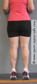
Ankle Rehab Exercises - Heel Raises
The calf muscles, including the gastroc and soleus are important during ankle function. They are the muscles that are responsible for you being able to point your toes, lift your heels up, walk, run, and jump. They also help control a lot of the movements that you perform during sports.

Strengthening of the calf can be accomplished with lots of different exercises. My favorite is the simple heel raise against the wall with forward support. 

Initially, you will want to do the heel raises on both feet at the same time, but as your ankle heals, you will be able to do single leg heel raises. 

Another way to strengthen the calf is with theraband exercises. Lunges, step ups, and balancing activities will also help to strengthen the calf muscles.

The Peroneals

The peroneal muscles are the stabilizers of the ankle. Located on the outside of the leg, they help the ankle maintain a neutral position when you are putting weight on your leg.

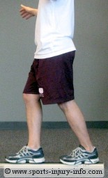
Try balancing on one foot for a few seconds. Watch what your ankle does while you are balancing.
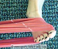
Peroneal Strengthening
Notice how your foot rolls in and out and adjusts to keep you balanced? The peroneal muscles help to keep your foot neutral while you are balancing, as well as when you are walking, running, jumping, and landing. 

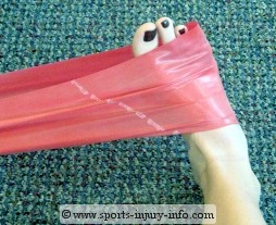
Strengthening of the peroneals can be done with theraband exercises. This type of ankle rehab exercise will isolate these muscles, and can be helpful in the early stages of your rehabilitation. 

Once you are stronger, more advanced exercises like lunges, step ups, and other balancing activities will help improve the peroneal's strength.

The Posterior Tibialis Tendon and Muscle

Posterior Tibialis Strengthening
The posterior tibialis is the medial stabilizing muscle of the calf. It helps to do the same thing as the peroneals, only on the opposite side. It works to keep the ankle and foot in a neutral position when you are bearing weight, and works with the peroneals to do this.

The posterior tibialis can also be strengthened with theraband exercises, and then more advanced exercises like lunges, step ups, and balancing.

Other Muscles

Functional Strengthening
There are several other muscles in and around the ankle that can be strengthened during ankle rehabilitation. The anterior tibialis, extensor digitorum, flexor digitorum, and other intrinsic muscles can all benefit from strengthening exercises.

It is important to remember though, that sports activities as well as daily life do not occur in isolation. So, ankle rehab exercises to improve your strength should also not occur in isolation.

In the early stages of rehab, some isolation is needed to retrain the muscles to be able to contract. This is very true if you have a lot of swelling. Theraband strengthening is a great way to work on isolated muscle activity.

After the first 1-2 weeks following initial rehabilitation, your program should progress to more multi-muscle exercises, like the lunges and step ups, and balancing activities discussed already. 

Ankle rehab exercises for improving strength should include the major muscle groups that produce the motions at the ankle as well as stabilize during activities. Your program should start out with simple exercises like theraband strengthening, and then progress to more advanced functional activities like lunges, step ups, and balancing.

Tuesday, October 23, 2012

Foreign bodies made in America


Most foreign bodies we see in our center are not incredibly too foreign. In fact, most of these objects tend to be naturally occurring debris from the oceans and lakes in Florida. Shells, stingray barbs, and even an occasional brackish water stone laceration to the plantar foot. If these injuries are not treated  in emergent fashion, this may lead to subsequent infection.  In the case above, this patient was running in a bonfire for some odd reason, and apparently one of the sharper wooden shards punctured the foot and simultaneously burned it, leaving him with second degree deep tissue burns. This was not immediately treated until 3 days later, and he ended up needing surgery to clean the wound and remove dead tissue within the area. He also requires IV antibiotics and several weeks of protected weight to the area with crutches until it all heals. We will see this during the "seasonal time" here mainly because more vacationing residents will be on the beaches, or wherever and they will not wear shoes. A foot injury like this is usually easily prevented just by wearing a sandal or sneaker at all times while outside. Most times, our feet are not well equipped to deal with the amount of sharp and dangerous objects that may lie waiting for unsuspecting tourists to walk on them. If you suspect you have stepped on something, don't wait. Come to see us at the FFLC, so we may rapidly diagnose, treat, and remove any foreign object from the foot before infection and need for hospitalization may occur. We can remove the foreign object right in office, and are well equipped to obtain cultures from the area, perform ultrasound guided removals, and even identify radio opaque material with use of radiography.

Tuesday, October 9, 2012

Green Bay Packers season in peril on account of a foot injury

The season looks dire so far in 2012 for any Green Bay fan, as first we lost a game on a call which was undoubtebly the most controversial and ridiculous call in NFL history to the Seahawks and now we lose to the Colts with a rookie quarterback and with that we also lose 3 more starters on both sides of the ball due to significant injuries. Not to mention that both the Bears and Vikings are winning every week now. We were 15-1 last year, and this year we probably will not make the playoffs as we are already 3rd place and have a 2 game deficit to both teams ahead of us with the Texans coming up next week who will likely demolish the Packers.

After losing to the otherwise rebuilding Colts, another unfortunate injury may have been partly responsible. The Packers have had a mediocre running game as it is this year with Benson, but now we have to struggle further without him. Maybe there is a good practice squad runner that nobody knows yet who can step it up and have a "next man up" mentality a la 2 years ago with our playoff run with an unknown running back. To make things even worse, with the loss of Cedric Benson, a running back who was supposed to be main addition to an offense that has had little trouble scoring points through the air last season, and give this team another dimension to confuse defenses and give Aaron Rodgers some help with the ground game, his ability to cut and balance and jump may have been permanently altered because of his foot injury. Teams will sit in zone defense and blitz packages knowing fully that we have no ground game to use and this will be a long year for certain.

Now, not only can we not pass the ball very consistently because the offensive line cannot block anyone, but we really will be left unable to run the ball as well, and Rodgers will continue to get sacked 5 or more times a game and potentially sustain another concussion, and we have nobody on reserve as a quarterback, because we let him go to Seattle in the offseason!! Fire Dom Capers too, because our defense cannot hold a three score lead against the COLTS!!

Anyhow...

Benson has suffered a midfoot sprain. This injury has been relatively common this year, with both Benson, and Santonio Holmes of the Jets suffering this injury already this year. Last year the Texans had a quarterback who also had a Lisfranc injury.
A most devastating injury, which can sideline a player for 2 months, and may lead to longterm pain and need for fusion if arthrosis and degenerative joint disease ensues. Football requires quick changes in direction, jumping, and running with quick stops and starts. All of these motions are difficult when a player has sustained such an injury. Longterm, we have noticed a number of patients who have likely had such an injury in the past and was not diagnosed or treated properly and are left with a change in the position and alignnment of the midfoot. This may lead to rockerbottom foot deformity in extreme cases, and most likely will lead to chronic recalcitrant pain in many cases. Ideally the bones should be reduced as soon as possible in cases of initial instability to their anatomic position, but this is often times not done on account of lacking evidence of fractures, or poorly obtained radiographs which do not show the alignment concerns from this injury secondary to the patient being unable to stand on the foot in order for the films to demonstrate the instability. Regardless, once the swelling has diminished with non weight bearing and casting, a flat film with the patient standing should be obtained to see if any instability has occured, otherwise the patient may be placed under anesthesia in order to use an active xray machine to stress the midfoot in order to demonstrate any need for fixation.

Monday, October 1, 2012

Turf the "Turf Toe" Injury


Often around this time of the year, football injuries become more prevelant in our practice with high school and junior leagues. The great toe will get hyper-extended for a brief moment, and this leads to pain and inability to return to sport. Sometimes this injury can lead to chronic instability of the toe joint which may lead to arthritis and eventual need for surgery if not treated properly with initial presentation. This injury is typically treated incorrectly with coritsone injections, and this may actually lead to further damage to the joint capsule and even the ligament between the sesamoid bones. Treatment for this condition starts with proper evaluation, detailed history and physical examination, and sometimes with foot blockade of local anesthetic or even arthrogram, we can diagnose and give a prognosis to return to the field in 1 visit. This treatment may consist of casting, immobilization, PRP injections, or even surgery for progressively more unstable joints or specific findings from MRI examination. Ultimately, athletes can expect a minimum of 2 weeks off, followed by gentle passive range of motion exercises, and finally we may allow return to the field for practice and eventual playing time. Most athletes will not return to the field if they are unable to dorsiflex the great toe joint at least 50 degrees without pain. If a patient has progressive deviation or valgus deformity becomes evident, surgery is most likely indicated. If the pain improves after proper immobilization and therapy driven rehabilitation, many will not require surgery.  Ultimately it is decided based on timely diagnosis, treatment, and therapy driven rehabilitation. Notable players who have had Turf Toe injury include Deion Sanders and Ray Lewis. Sometimes this injury will end the professional player's career.

Monday, September 17, 2012

Stem Cells and Placental Growth Factors

Often times, new technology is encountered in any medical subspecialty that raises eyebrows and concerns patients. This is usually met with skepticism and may even offer need for second opinions depending on the nature of the treatment in question.  This practice is always striving to be on the cutting edge of new technology and we have always tried to offer all nonsurgical management options possible depending on the clinical pathology at hand. We see many patients each day with chronic tendon tears, or capsular injuries which are subtle, and most often longstanding. These sorts of injuries range from mild symptoms, to severe tendon pathologic weakness. Often times these injuries and chronic conditions would require bracing and surgery, but more biotechnology is becoming available with new data suggesting alternatives to these sorts of treatments. There are several companies which are bringing cellular technology that contains growth factors such as PDGT A and B, FGF, TGF-beta, EGF, and various others with stem cell progenitors which may lead to proliferating tissues in vitro.  This is exciting, primarily for patients who otherwise are told they need extensive tendon debridement and reinforcements, otherwise they require extensive bracing and immobilization which will limit activity level and lead to further health issues longterm.  This technology may not heal a tendon which is 80% torn, or increase range of motion in a great toe joint with capsular damage that has led to cartilage compromise, but it may be the sort of technologic treatment to serve as another intermediate step in the progressive nature of treating chronic foot and ankle tendonopathys and capsulopathies which may prolong or negate the time frame to need any sort of surgery.  Data is still being amassed and certainly no final consensus has been reached as to how much these sorts of injectable treatments can benefit patients in differing tendon dysfunctions, but one thing is certain: FFLC will be there to at least help evaluate these treatments and offer all beneficial treatments possible to our patients.

DR TIMM

Wednesday, August 29, 2012

NFL reciever Nicks may return to line up tonight.


One of the more prolific pass catchers for the NFL New York Football Giants will return to his number 88 jersey possibly tonight after recovering from his fifth metatarsal fracture ORIF surgery from May 2012. He likely had a Jones fracture, which in most cases with athletic players a percutaneous screw was utilized in order to fascilitate healing. At FFLC, you are not required to be an NFL player to get this state of the art procedure if necessary, as we have successfully treated numerous athletic patients with this traumatic injury using this specific technique with minimal to no complications.

The main reason why surgery is likely required in these individuals with the fractured fifth metatarsal, stems from the nature of the amount of weight applied to the area with each step, as well as where the tendons insert at the most proximal aspect of the main fracture fragment, with each step the tendon pulls away this fracture from the main portion of the bone.  Sometimes a below knee cast is applied to stop all motion, but even this may not fully accelerate healing. The screw fixation eliminates all motion to the site, and by placing the screw, the surgeon is able to drill through the scar tissue and promote healing with placement of demineralized bone matrix proteins and collagen to enhance liklihood of healing the fracture.

So if you happen to catch the Patriots and Giants tonight, keep your eyes on number 88, as his cutting and route running is still going to be at a high level after his injury, because he had state of the art surgery in order to heal his fracture.

Tuesday, August 21, 2012

Chemotherapy and your feet.

Some of our patients are undergoing chemotherapy to treat various cancers, and this may cause some patients to experience painful scaling skin on the palms and soles of their feet and hands. Here is some general information to guide you towards improving this condition. Please come and see me if any problems or concerns arrise.

- Dr Timm

Hand-Foot Syndrome
Other terms:
Palmar-Plantar Erythrodysesthesia; PPE What is hand-foot syndrome?Also called hand-foot syndrome or hand-to-foot syndrome, Palmar-Plantar Erythrodysesthesia is a side effect, which can occur with several types of chemotherapy or biologic therapy drugs used to treat cancer. For example, Capecitabine (Xeloda®), 5-Flurouracil (5FU), continuous-infusion doxorubicin, doxorubicin liposomal (Doxil®), and high-dose Interleukin-2 can cause this skin reaction for some patients. Following administration of chemotherapy, small amounts of drug leak out of very small blood vessels called capillaries in the palms of the hands and soles of the feet. Exposure of your hands and feet to heat as well as friction on your palms and soles increases the amount of drug in the capillaries and increases the amount of drug leakage. This leakage of drug results in redness, tenderness, and possibly peeling of the palms and soles. The redness, also known as palmar-plantar erythema, looks like sunburn. The areas affected can become dry and peel, with numbness or tingling developing. Hand-foot syndrome can be uncomfortable and can interfere with your ability to carry out normal activities.
Things you can do if you suspect hand-foot syndrome (Palmar-Plantar Erythrodysesthesia):
Prevention:
Prevention is very important in trying to reduce the development of hand-foot syndrome. Actions taken to prevent hand-foot syndrome will help reduce the severity of symptoms should they develop.This involves modifying some of your normal daily activities to reduce friction and heat exposure to your hands and feet for a period of time following treatment (approximately one week after IV medication, much as possible during the time you are taking oral (by mouth) medication such as capcitabine). Avoid long exposure of hands and feet to hot water such as washing dishes, long showers, or tub baths. Short showers in tepid water will reduce exposure of the soles of your feet to the drug. Dishwashing gloves should not be worn, as the rubber will hold heat against your palms. Avoid increased pressure on the soles of the feet or palms of hands. No jogging, aerobics, power walking, jumping - avoid long days of walking. You should also avoid using garden tools, household tools such as screwdrivers, and other tasks where you are squeezing your hand on a hard surface. Using knives to chop food may also cause excessive pressure and friction on your palms.  Cooling procedures:
Cold may provide temporary relief for pain and tenderness caused by hand-foot syndrome.
Placing the palms or bottoms of your feet on an ice pack or a bag of frozen peas may be very comforting. Alternate on and off for 15-20 minutes at a time.
Lotions:
Rubbing lotion on your palms and soles should be avoided during the same period, although keeping these areas moist is very important between treatments.
Emollients such as AMLACTIN, Aluvea,Aveeno®, Lubriderm®, Udder Cream®, and Bag Balm® provide excellent moisturizing to your hands and feet.
Pain relief:
Over the counter pain relievers such as acetaminophen (Tylenol®) may be helpful to relieve discomfort associated with hand-foot syndrome. Check with your doctor.
Vitamins:
Taking Vitamin B6 (pyridoxine) may be beneficial to preventing and treating Plantar-Palmar Erythrodysesthesia, and should be discussed with your doctor.
Drugs/treatment changes that may be prescribed by your doctor:
Chemotherapy treatments may need to be interrupted or the dose adjusted to prevent worsening of hand-foot syndrome.
When to call your doctor or health care professional:
If you notice that your palms or soles become red or tender. This most often occurs before any peeling, and recommendations for relief of discomfort can be given. If you are on chemotherapy pills, you may be asked to hold treatment, or need your dose adjusted to prevent worsening of symptoms.

Friday, August 10, 2012

Pain in the Achilles !!

So many of our patients suffer from what we in the medical field call "Tendonopathy". This is a vague term which encompasses a vast array of varying pathologic conditions, in both acute and chronic presentations.

Probably one of the main contributers to the more common tendonopathy that we see is the pressence of a large spur behind the heel which is associated near the insertion of the most important tendons in the foot and ankle - ie the Achilles tendon.

Numerous treatments in the literature have been described, and the main thing to consider in these situations, is that there are viable nonsurgical options available for this disorder. Many patients hesitate to present in the office of a physician with lumps and bumps on their extremities for fear of needing surgery. This is a problem, because most of the time these anxieties allow the pathologic process to worsen, and eventually by delaying treatment you are nearly obligating yourself to undergo the one treatment you were trying to avoid.  Surgery is always a last option for posterior heel pain, as this can require 3 months or more to recover from depending on the nature of the pathology and extent of procedures required to rectify the conditions. Many times, we can identify the extent of the pathology and at least rule out tearing or masses with the use of ultrasound technology which is readily available in the office. Often, we require MRI images to plan should nonsurgical options fail to reduce symptoms.

We offer state of the art high energy shock wave therapy coupled with PRP injections to the posterior achilles for pain and thickening, and to reduce symptoms from protruding and prominent boney areas behind the heel that may be penetrating through the tendon in this part of the foot, thus causing tendonopathy pain. This treatment is more effective in less chronic cases, but is usually able to reduce the pain and edema from these conditions in most cases. Shoegear choices are also a factor here, as many ill fitting shoes may contribute to pain in areas of boney prominences.

Do not hesitate to make an appointment, as we may be able to reduce your pain without surgery !

Monday, July 30, 2012

Calcinosis ?


Here at FFLC, we are asked to see a multitude of varied pathologies by surrounding physicians, and we will on occasion recieve difficult wounds which are otherwise well treated, but still fail to heal. This is something that may require advanced care and a more thorough diagnostic approach.

Sometimes wounds will not heal for more obvious reasons, such as too much pressure on the area or underlying bone infections that are present which will preclude closure of the site. But we will also see underlying metabolic pathology which can go unrecognized and lead to failure of wound healing; such as in the case of underlying calcinosis cutis.

What is this?

This can be seen sometimes on plain radiographic images, where one may see some abnormality in the soft tissue which can look like a bone, or bone densities within the soft tissue. This is somewhat rare, but may lead to wound healing delays, and even pain.

Other times this may be found only on skin biopsies of the ulcer may find the calcifications on a microscopic level, and in my opinion any peculiar skin lesion should have a biopsy done to check for cancer or any other pathology that may otherwise not be detected.  In fact, in the case of any non healing ulcer that is present for more than 4 weeks, a biopsy should always be done to further assess and treat the site along with bacterial culture and even CBC and C-reactive protein with BMP labwork.

Basically, there are varieties of presentations of calcinosis cutis, but the main concept here is that it may cause wound healing delays or preclude any healing from the site. This may require systemic treatment and evaluation from renal specialists, rheumatology specialists, and even vascular surgeons to some extent. Regardless, extensive workup must be entertained in any wound care patient in order to identify reasons for nonhealing. This also potentiates the need for aggressive debridement, as the tissue itself may be too compromised for any reasonable healing to occur, and with an aggressive debridement the tissue may be allowed to "reset" and build the scaffolding for dermal/epidermal cells again from "scratch".


Monday, July 16, 2012

OSTEOPOROSIS is a real risk for bone healing.


Two major factors that influence the risk of development of
osteoporosis are the level of bone mass achieved at skeletal
maturity (peak bone mass) and the rate at which bone loss
occurs in later years. The more bone mass available before
age-related bone loss ensues, the less likely it will decrease to
a level at which fractures occur.
Research studies point to a number of risk factors that
may have a strong influence on peak bone mass and the rate
of bone loss, and thus the development of osteoporosis
Some of these factors include: inadequate nutritional
intake, lack of physical activity, smoking, excessive alcohol
consumption, and prolonged use of corticosteroids.
In addition to diet and lifestyle factors, genetic and
ethnic factors significantly influence many aspects of
calcium and skeletal metabolism. Caucasian and Asian
women tend to have lower bone density than African and
Hispanic women and, consequently, are more likely to suffer
from osteoporotic fractures. The same holds true for thin,
smaller boned women. Evidence also suggests there may
be a link between mother and daughter; mothers with
low bone mineral content tend to have daughters with low
bone mineral content. Whether this link is a function of
heredity or the influence of the mother’s habits, or both,
remains uncertain.

HOW MUCH AND WHAT DO WE NEED FOR OUR BONES?

The Recommended Daily Allowance (RDA) for

calcium is currently set at 800 mg for individuals 1-10 years
old and 25 years and older and 1,200 mg for those 11-24
years old and for pregnant or lactating women. However,
these levels are well below the level of intake that many
experts recommend. The authors of a study of recent
intervention trials of calcium supplementation recommended that
the RDA during childhood should be 1,250 mg and 1,450 mg
during adolescence, while others have recommended a
calcium intake of up to 1,800 mg/day during adolescence.
Such an increase in calcium intake during adolescence could
play an important role in the attainment of optimal peak
bone mass.
Regarding the calcium intake for older individuals, many
experts recommend an intake of 1,500 to 2,000 mg/day to
minimize bone loss in some patients. The National
Institutes of Health (NIH) Consensus Conference on Optimal
Calcium Intake recommends calcium intakes of 1,200 to
1,500 mg for 11-24 year olds, 1,000 mg for those 25-50 years,
and 1,500 mg for those over 65.3 In addition, the NIH recommends
a calcium intake of 1,500 mg/day for women over 50
years who are not receiving hormone replacement.
While the RDA levels of calcium may be a source of
debate, the real issue is the fact that a large proportion of the
population isn’t even meeting the current RDA
levels. According to data obtained from the USDA’s
1987-88 Nationwide Food Consumption Survey, the mean
per capita daily consumption of calcium for the U.S.
population was 737 mg. The data for women as a group was
even worse: after age 11, no age group of females achieved
even 75% of the RDA for calcium. And between the ages of
12 to 29, when calcium requirements reach their peak because
of rapid skeletal growth, women consumed <60% of the RDA
for calcium. Therefore, the challenge for the health-care
professional is to educate patients on the importance of lifetime
maintenance of adequate calcium intake.

VITAMIN D


Vitamin D plays an essential role in maintaining a
healthy mineralized skeleton. The main physiologic function
of vitamin D is to maintain serum calcium and phosphorus
concentrations within the normal range to maintain essential
cellular functions and to promote mineralization of the
skeleton. Vitamin D acts primarily to increase serum
calcium by stimulating intestinal absorption of calcium.
Vitamin D insufficiency results in reduced calcium absorption,
a rise in circulating parathyroid hormone, and increased bone
resorption. The elderly often have a low level of vitamin D
deficiency owing to less efficient skin synthesis of vitamin D,
less efficient intestinal absorption, and reduced sun exposure
and vitamin D intake.

Vitamin D deficiency can result in secondary
hyperparathyroidism, a condition that accelerates bone
resorption and thus exacerbates osteoporosis. Vitamin D
deficiency is associated with increased risk of hip fracture,
and several studies have demonstrated that an increase in
calcium intake of 800-1000 mg/day with supplementation of
400-800 units of vitamin D daily will decrease the risk of
vertebral and nonvertebral fractures and increase bone
mineral density.

MAGNESIUM

Although decreased bone mass is the hallmark of
osteoporosis, qualitative changes in bone matrix are also
present, which could result in fragile or brittle bones that are
more susceptible to fracture. There is growing evidence that
magnesium may be an important factor in the qualitative
changes of the bone matrix that determine bone fragility.
Magnesium influences both matrix and mineral metabolism in
bone by a combination of effects on hormones and other
factors that regulate skeletal and mineral metabolism, and by
direct effects on bone itself. Magnesium depletion affects all
stages of skeletal metabolism adversely, causing cessation of
bone growth, decreased osteoblastic and osteoclastic activity,
osteopenia, and bone fragility.

SUPPLEMENTATIONS

Microcrystalline hydroxyapatite concentrate (MCHC) is

an excellent source of bioavailable calcium.27,53-56 MCHC is
derived from whole bone and is complete with the minerals
and organic matrix found in raw bone. In addition to calcium
and the organic components (mostly collagen protein and
mucopolysaccharides), MCHC contains phosphorus,
magnesium, fluoride, zinc, silicon, manganese, and other
trace minerals in the same physiological proportions found in
healthy bone.

Talk to your endocrinologist or primary health care provider
if you are osteoporotic and they may need to prescribe medications
as well to increase the density of your bones to prevent fractures,
improve bone healing potential, and allow for more active and
healthy lifestyles to be preserved.

Tuesday, July 10, 2012


Smoking is a major problem:


More than 1 billion people worldwide smoke and annually, 5.4 million deaths or 1 death every 6 seconds, is due to tobacco smoking. The prevalence of cigarette smoking in England is at a record low with 21% adult smokers in 2008. Nevertheless, smoking is attributable to 80,000 deaths per year in England and an estimated 8.5 million people still smoke in the United Kingdom.

It is now well known that cigarette smoking has adverse effects on the human body, notably its association with lung cancer.

Anecdotal experiences associating wound healing complications with smoking are well documented in the literature. Despite this, there are only a few large clinical trials attempting to elucidate the relationship between tobacco smoking and wound healing. This has resounding implications not only on the surgical patient, the surgeon but the economics of the NHS as well. The repercussions of abnormal wound healing are perhaps best felt in plastic surgery and in podiatry, where aesthetic value is as important as functional value.

A good portion of patients that we perform surgery on are undergoing elective procedures for reduction in pain, ease of fitting in shoes, and to a lesser degree small digit cosmesis to realign a toe or toes. This is imperitive to understand that as the vessels branch from the popliteal fossa to medium and small named vessels, they become even smaller passed the midtarsal joint into the toes. This directly is impacted by the mechanism which nicotine and other chemicals that are found in cigarrette smoke directly and indirectly lead to vasoconstriction which pinches closed the supply to the tissues which have been traumatized by surgery. This leads to delayed wound healing time and thickened scars, and may lead to gangrene and amputation in select cases.

Effects of smoking at a cellular level


There are more than 4,000 chemicals present in a cigarette. Amongst these chemicals, nicotine and carbon monoxide are important contributors to the detrimental effects smoking has on wound healing.

Carbon monoxide has a 200 times greater affinity to haemoglobin than oxygen. This results in tissue hypoxia as the oxygen-haemoglobin saturation curve shifts to the left. Consequently, the wound healing process is impaired due to the hypoxic state in tissues.

Nicotine acts on the dermal-subcutaneous vascular plexus to cause vasoconstriction. This has serious consequences as many random patterned flaps rely heavily on the plexus for survival. Besides inducing a hypoxic state and causing vasoconstriction, smoking also leads to increased platelet aggregation, creating microangiopathic thromboses which are tiny blood clots in the capillaries. This is particularly detrimental at the wound site as healing depends heavily on existing and new capillaries.

High levels of fibrinogen and haemoglobin are found in smokers and coupled with reduced fibrinolytic activity, local perfusion to the wound site is greatly attenuated, resulting in delayed wound healing.

This is supported by the work of Sarin et al who found that smoking one cigarette could reduce blood velocity by 42%.

In summary, if you smoke, you may be greatly compromising the chances your elective surgery will not be without complications.
 
Here is a published algorithm which may answer why you may be denied elective surgery if you smoke, because it is in your best interests:


Wednesday, June 20, 2012


Drinking Enough Water?

Hydration is a primary key to performance in sports, especially in the case of high endurance requiring activity like running, playing basketball, soccer, or tennis. We see a number of patients who are coming in with chronic aching legs and "charlie horse" variety of symptoms at night. These patients are not typically reporting any specific injury, but they are stating in general that sleep is difficult, and "locking" or "knotting" in the muscles is noted. These typically lead to immediate need to discontinue current activity, and keep area extended to prevent flexion locking while actively experiencing the cramping sensations.

If you've ever had muscle spasms or muscle cramps, you know they can be extremely painful. In some cases, a muscle may spasm so forcefully that it results in a bruise on the skin. Most muscle spasms and cramps are involuntary contractions of a muscle. A serious muscle spasm doesn't release on its own and requires manual stretching to help relax and lengthen the shortened muscle. Spasms and cramps can be mild or extremely painful. While they can happen to any skeletal muscle, they are most common in the legs and feet and muscles that cross two joints (the calf muscle, for example). Cramps can involve part of a muscle or all the muscles in a group. The most commonly affected muscle groups are:


•Back of lower leg / calf (gastrocnemius).

•Back of thigh (hamstrings).

•Front of thigh (quadriceps).

•Feet, hands, arms, abdomen

Muscle cramps range in intensity from a slight twitch or tic to severe pain. A cramped muscle can feel rock-hard and last a few seconds to several minutes or longer. It is not uncommon for cramps to ease up and then return several times before they go away entirely.


What Causes Muscle Cramps

The exact cause of muscle cramps is still unknown, but the theories most commonly cited include:

•Altered neuromuscular control

•Dehydration

•Electrolyte depletion

•Poor conditioning

•Muscle fatigue

•Doing a new activity

Other factors that have been associated with muscle cramps include exercising in extreme heat. The belief is that muscle cramps are more common during exercise in the heat because sweat contains fluids as well as electrolyte (salt, potassium, magnesium and calcium). When these nutrients fall to certain levels, the incidence of muscle spasms increases. Because athletes are more likely to get cramps in the preseason, near the end of (or the night after) intense or prolonged exercise, some feel that a lack of conditioning results in cramps.


Research Supports Altered Neuromuscular Control as the Cause of Cramps

While all these theories are being studied, researchers are finding more evidence that the "altered neuromuscular control" hypothesis is the principal pathophysiological mechanism the leads to exercise-associated muscle cramping (EAMC). Altered neuromuscular control is often related to muscle fatigue and results in a disruption of muscle coordination and control.

According to a review of the literature conducted by Martin Schwellnus from the University of Cape Town, the evidence supporting both the "electrolyte depletion" and "dehydration" hypotheses as the cause of muscle cramps is not convincing. He reviewed the available literature supporting these theories and found mostly anecdotal clinical observations and one small case-control study with only 10 subjects. He also found another four clinical prospective cohort studies that clearly did not support the "electrolyte depletion" and "dehydration" hypotheses as the cause of muscle cramps. In his review, Schwellnus concludes that the "electrolyte depletion" and "dehydration" hypotheses do not offer plausible pathophysiological mechanisms with supporting scientific evidence that could adequately explain the clinical presentation and management of exercise-associated muscle cramping.

"Scientific evidence for the "altered neuromuscular control" hypothesis is based on evidence from research studies in human models of muscle cramping, epidemiological studies in cramping athletes, and animal experimental data. Whilst it is clear that further evidence to support the "altered neuromuscular control" hypothesis is also required, research data are accumulating that support this as the principal pathophysiological mechanism for the aetiology of exercise-associated muscle cramping (EAMC)."

Treating Muscle Cramps

Cramps usually go away on their own without treatment, but these tips appears to help speed the healing process:

•Stop the activity that caused the cramp.

•Gently stretch and massage the cramping muscle.

•Hold the joint in a stretched position until the cramp stops.

Preventing Muscle Cramps

Until we learn the exact cause of muscle cramps, it will be difficult to say with any confidence how to prevent them. However, these tips are most recommended by experts and athletes alike:

•Improve fitness and avoid muscle fatigue

•Stretch regularly after exercise

•Warm up before exercise

•Stretch the calf muscle: In a standing lunge with both feet pointed forward, straighten the rear leg.

•Stretch the hamstring muscle: Sit with one leg folded in and the other straight out, foot upright and toes and ankle relaxed. Lean forward slightly, touch foot of straightened leg. (Repeat with opposite leg.)

•Stretch the quadriceps muscle: While standing, hold top of foot with opposite hand and gently pull heel toward buttocks. (Repeat with opposite leg.)

Most muscle cramps are not serious. If your muscle cramps are severe, frequent, constant or of concern, see your doctor.


Source http://sportsmedicine.about.com/od/legpainandinjuries/a/muscle_spasms.htm

Monday, April 2, 2012

The Mini Bunion

"I have a bunion on the outside of my foot"

This is more commonly called a bunionette, which is a small bunion occurring on the outside of the foot. They are also called tailor’s bunions, which likely stemmed from a shoemaker's peril in constructing fitting shoes for patients with extremely wide distal feet. Regular bunions affect the big toe, while a bunionette is a bunion on the pinky toes. A bunionette is a visible bump along the outside of the foot at the base of the little toe.During the development of a bunionette, the little toe is being pushed over towards the other toes. Over time, this causes the joint at the base of the little toe to protrude. Without treatment, bunions and bunionettes may progress to a completely dislocated toe with excessive widening of the angle between the fourth and fifth metatarsal bones. A painful callous, "Lister's" corn, or other abnormality may form at the site of the bunionette.Tailor’s bunions often happen to people who have feet which pronate excessively during their gait. These smaller bunions can also be a result of footwear that doesn’t fit properly. A more uncommon cause may be a genetic predisposition towards bunions which is passed from parents to offspring in a seemingly predictable pattern. A doctor will rule out other causes for the pain and swelling, such as arthritis or joint infection.Another cause, as the pseudonym “tailor’s bunion” implies, could be related to occupational or habitual pressure on the outside of the foot. The name comes from the fact that tailor’s were prone to these small, external bunions due to the position they kept their feet in while working, which may have caused pressure to the joint leading to bursal formation and eventual lateral capsule "wear and tear" leading to the medial pull of the toe towards the others.Though it is a possible cause, bunionettes are almost always caused by footwear. Ballet dancers are prone to bunionettes due to the tight ballet shoes.Bunionettes can be painful, especially when irritated by friction from footwear. A non-surgical treatment option for a bunionette involves buying shoes that allow more room for the toes. This may be coupled with various cushions and even arch supportive orthotics to reduce the abnormal pronation which may allow for more rapid progression of the deformity.Someone with tailor’s bunions may choose to go barefoot or wear sandals to stop this irritation. A shoe repair shop may be able to stretch shoes that are uncomfortable. In some cases, wearing footwear that doesn’t put pressure on the foot is the only treatment needed.Using nonsteroidal anti-inflammatory drugs (NSAIDS) may help relieve any pain or discomfort. Elevating the foot and applying ice to the affected area can give some relief. Any bunions occurring in children require medical attention.Using bunionette pads can help alleviate the pain. A bunionette pad cushions the affected area and stops friction from footwear. Bunionette pads are available at most drug stores. Moleskin or felt patches can be used to stop the bunionette from rubbing against the shoes.Tailor’s bunion surgery is an option if the bunionette is problematic. Bunion foot surgery should only be considered if the non-surgical bunion treatments fail to provide relief. The surgeon will order tailor’s bunion x ray to check the degree of damage to the foot. The surgeon will consider this and other factors to decide which procedure or procedures will be most effective to relieve your symptoms.

Monday, March 12, 2012

Season in full swing

Every year during when the weather is cold "up north" the weather stays nice here in Naples, and so the patient volume can triple secondary to seasonal residents seeking foot and ankle care in our center. This time seems hectic and many patients are trying to enjoy the time that they share between their northern homes and their vacation homes in this area. This can sometimes be hampered by injury, sickness, and even unforseen foot and ankle pain. At our center, we strive to reduce pains from tendonitis, relieve infections from insect bites and stingray punctures, and take care of any other short or long term concern which just will not succumb to the schedules of our patients during their vacations in Naples. We are constantly treating patients during their stay in our area and even if this is strictly temporary until they return North, we are able to guarantee excellent care by means of extensive training as well as by extensive experience.

Irregardless the size of the injury or extent of the infection, we will keep you safe until you can get home in time for the summer. With our electronic records system, we can even email records to a physician to fascilitate ease of transition from here to home. And we are glad to follow up with any concerns which may carry over from home to here as well. Keep that in mind as you walk the beachlines, or play tennis and golf and thing are just not feeling quite right. There is always enough time to get you at least on track towards enjoying the rest of your vacation if you are treated at FFLC.

Wednesday, January 11, 2012

New Years Resolutions

Every year around mid January and into February, we get an influx of patients who are very driven to lose weight secondary to overindulgent eating during the holiday season. This leads to a high degree of previously inactive individuals who are developing heel pain from their newly achieved activity level. This creates a formidable foe with regards to maintaining an exercise level consistant with weight loss goals.

At FFLC we strive to get you back out there running, and this is done via a variety of measures for treatment of heel pain, which consists of well conforming inserts for your shoes, as well as ultrasound guided examinations to pinpoint pathology and isolate sore bursal formations and fascia tears. We strive to keep this community active, and usually this can be managed without surgical interventions. We routinely perform physician guided physical therapy at out PT office and for difficult to resolve pain, we will utilize state of the art extracorporal shock wave therapy.

When you are not stretching your legs and heels appropriately, a night splint may be very useful to allow you to stretch out the fascia and reduce recurrence of painful heels. With xrays on site and experienced foot and ankle surgeons on your side, it is no wonder Naples continues to stay active, even when heel pain becomes part of the equation.